. 2007 Jul-Aug;32(7-8):595-609.
doi: 10.1080/02713680701446653.

Ocular surface expression and in vitro activity of antimicrobial peptides

Affiliations

Ocular surface expression and in vitro activity of antimicrobial peptides

Ling C Huang et al. Curr Eye Res. 2007 Jul-Aug.

Abstract

Purpose: Human ocular surface epithelia express four antimicrobial peptides (APs): beta -defensin (hBD) 1-3 and LL-37. Here the expression of additional APs (hBD 4-6, HE2beta 1; histatin-1, -3; liver expressed antimicrobial peptide-1, -2; macrophage inflammatory protein (MIP)-3alpha, and thymosin (T)beta -4) was sought and activity against common ocular pathogens studied.

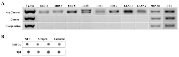
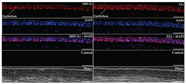
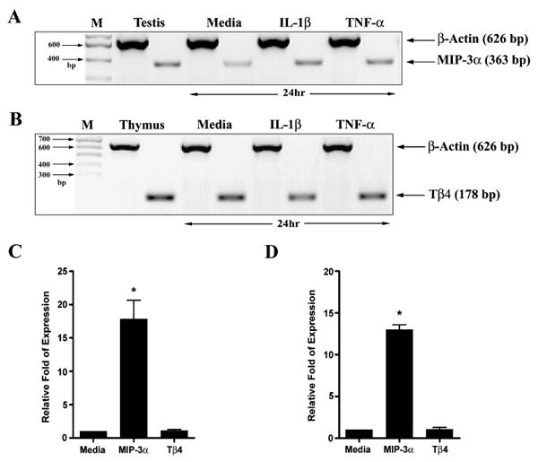
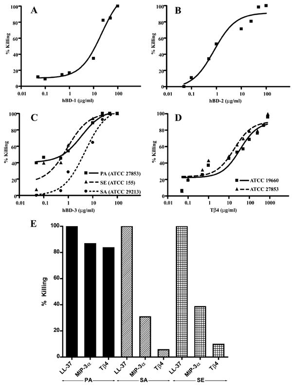
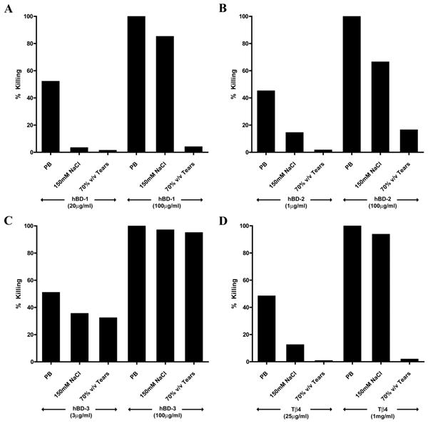
Methods: AP expression was determined in human corneal and conjunctival epithelial cells (HCEC, HCjEC) by RT-PCR and in corneal sections by immunostaining. Antimicrobial assays were performed to assess peptide (hBD 1-3, LL-37, MIP-3alpha, and Tbeta 4) activity against Pseudomonas aeruginosa (PA), Staphylococcus aureus (SA), and Staphylococcus epidermidis (SE) in the presence of NaCl or tears.

Results: HCEC and HCjEC expressed MIP-3alpha and Tbeta 4. hBD 1-3, MIP-3alpha, and Tbeta 4 showed activity against PA. hBD-3 had potent activity against SA and SE, whereas hBD-2, MIP-3alpha and Tbeta 4 had moderate activity and hBD-1 had none. NaCl markedly attenuated, and tears almost completely inhibited the activity of hBD 1-2 and Tbeta 4, but not that of hBD-3.

Conclusions: The ocular surface epithelia additionally express MIP-3alpha and Tbeta 4 which have moderate antimicrobial activity. The current data support a role for hBD-3 as an antimicrobial peptide in vivo, but call in to question the effectiveness of some other APs. However, further study is required to conclusively elucidate the physiological role of each AP.


Figures

Figure 1
Figure 1
Expression of MIP-3α and Tβ4 mRNA and protein by ocular surface epithelia. (A) RT-PCR. The figure shows representative results for cornea = scraped human corneal epithelium (n = 3); conjunctiva = primary cultured human conjunctival epithelial cells (n = 2); + ve controls = positive controls: testis (hBD 4–6, HE2β1, MIP-3α), salivary gland (Hist-1, -3), liver (LEAP 1-2), and thymus (Tβ4). (B) Immunoblotting. The figure shows representative results for: STD (standard) = 5 ng human rMIP-3α or 10 ng Tβ4 synthetic peptide; scraped = 25 μg cellular protein from two scraped human corneal epithelial samples (n = 2); cultured = 25 μg cellular protein from primary cultured human corneal epithelial cells (n = 3).
Figure 2
Figure 2
Immunostaining of MIP-3α and Tβ4 in the cornea. The figure shows representative images for MIP-3α (left) and Tβ4 (right) expression in normal human corneal epithelium (sections from central cornea). Specific immunolabeling for MIP-3α or Tβ4 is shown in red; DAPI: nuclear labeling (blue); MIP-3α or Tβ4+ DAPI: merged image; control: background labeling in the absence of primary antibody; phase: phase contrast image. Identical findings were seen in corneas from three different donors. (Scale bars: 40 μm).
Figure 3
Figure 3
Effect of pro-inflammatory cytokines on MIP-3α and Tβ4 expression in ocular surface epithelial cells. MIP-3α (A) and Tβ4 (B) mRNA expression by corneal epithelial cells treated with IL-1β(10 ng/ml) or TNF-α (10 ng/ml) for 24 hours. M = base pair marker. Real-time PCR showing relative levels of mRNA expression for MIP-3α and Tβ4 in IL-1β (C) or TNF-α (D) treated epithelial cell samples. The figure shows representative results for primary cultured HCEC (n = 3, * p < 0.05, compared to the untreated control). Media = normalized expression in media-treated samples set to one.
Figure 4
Figure 4
Antibacterial effect of hBD 1-3 and Tβ4 against common ocular pathogens. (A-B) 107 cfu/ml PA (ATCC 27853) were incubated with hBD-1 (A) or hBD-2 (B) for 2 hours. The graphs show the average data of three experiments. The EC50values were 21.4 ± 1.5 and 1.2 ± 0.2 μg/ml for hBD-1 and hBD-2, respectively. (C) 107 cfu/ml PA (ATCC 27853), SA (ATCC 29213), and SE (ATCC 155) were incubated with hBD-3 for 2 hours. The graph shows the average data of three experiments against each strain. The EC50values were 3.4 ± 1.3 (PA), 5.3 ± 1.5 (SA), and 0.9 ± 0.2 μg/ml (SE). (D) 107 cfu/ml PA (ATCC 27853 and ATCC 19660) were incubated with Tβ4 for 2 hours. The graph shows the average data of three experiments against each strain. The EC50 values were 18.4 ± 1.5 μg/ml (PA, ATCC 27853) and 28.7 ± 1.6 μg/ml (PA, ATCC 19660). (E) 107 cfu/ml PA (ATCC 27853), SA (ATCC 29213), and SE (ATCC 155) were incubated with LL-37 (50 μg/ml), MIP-3α (25 μg/ml), and Tβ4 (200 μg/ml) at 37°C for 2 hours. The graph shows representative data from two experiments.
Figure 5
Figure 5
Antibacterial effect of hBD 1–3 and Tβ4 against Pseudomonas aeruginosa (ATCC 27853) in the presence of NaCl and Tears. 107 cfu/ml PA were incubated with (A) hBD-1 (20 μg/ml or 100 μg/ml), (B) hBD-2 (1 μg/ml or 100 μg/ml), (C) hBD-3 (3 μg/ml or 100 μg/ml), or (D) Tβ4 (25 μg/ml or 1 mg/ml) in the presence or absence of 150 mM NaCl or tears (70% v/v) at 37°C for 2 hours. The graph shows representative data from one of two or three experiments. PB = phosphate buffer, control.

Similar articles

Cited by

References

    1. Sack RA, Nunes I, Beaton A, et al. Host-defense mechanism of the ocular surfaces. Biosci Rep. 2001;21:463–480. - PubMed
    1. McDermott AM. Defensins and other antimicrobial peptides at the ocular surface. The Ocular Surface. 2004;2:229–247. - PMC - PubMed
    1. Patrzykat A, Douglas SE. Antimicrobial peptides: cooperative approaches to protection. Protein Pept Lett. 2005;12:19–25. - PubMed
    1. Beisswenger C, Bals R. Functions of antimicrobial peptides in host defense and immunity. Curr Protein Pept Sci. 2005;6:255–264. - PubMed
    1. McClellan KA. Mucosal defense of the outer eye. Surv Ophthalmol. 1997;42:233–246. - PubMed

Publication types

MeSH terms

LinkOut - more resources