. 2007 Oct 3;26(19):4189-202.
doi: 10.1038/sj.emboj.7601852. Epub 2007 Sep 13.

Direct stimulation of receptor-controlled phospholipase D1 by phospho-cofilin

Affiliations

Direct stimulation of receptor-controlled phospholipase D1 by phospho-cofilin

Li Han et al. EMBO J. .

Abstract

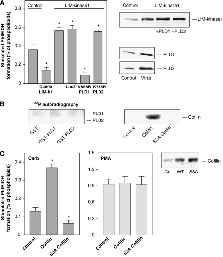
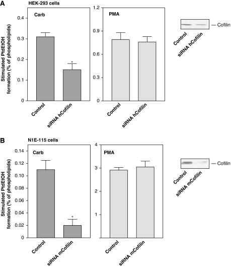
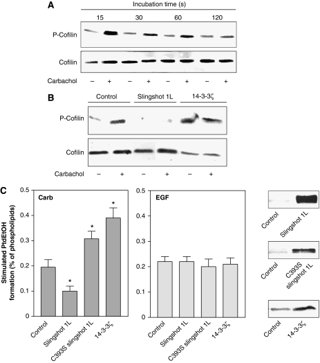
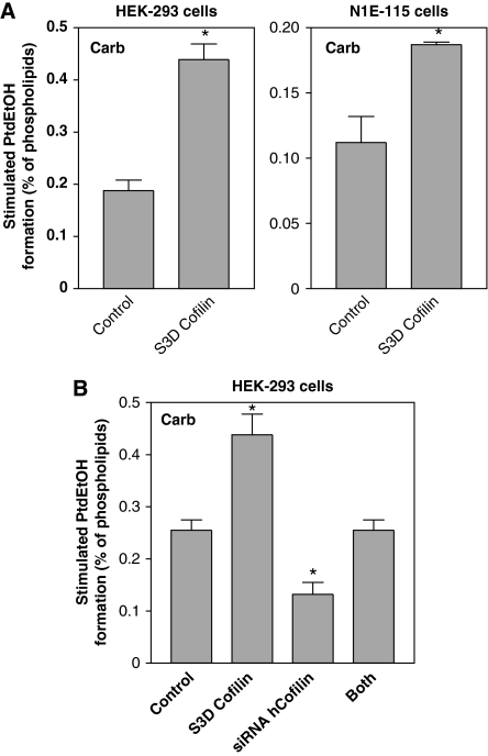
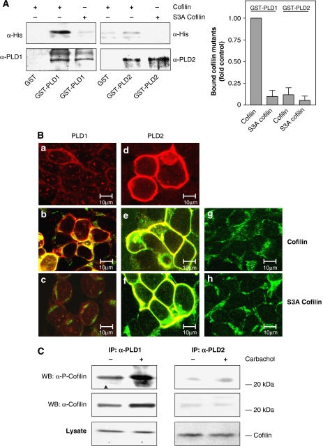
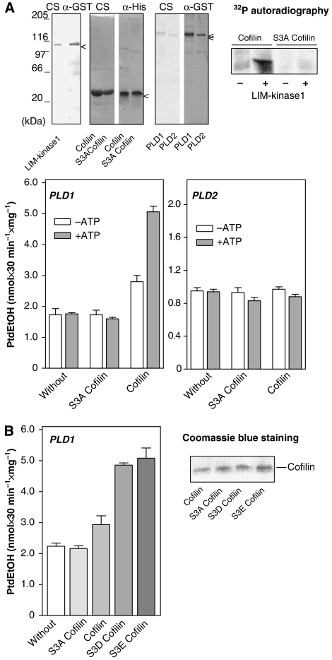
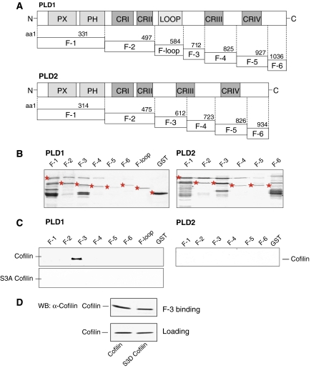
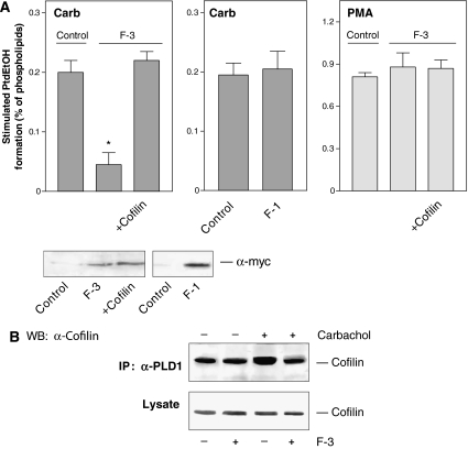
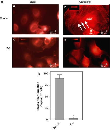
The activity state of cofilin, which controls actin dynamics, is driven by a phosphorylation-dephosphorylation cycle. Phosphorylation of cofilin by LIM-kinases results in its inactivation, a process supported by 14-3-3zeta and reversed by dephosphorylation by slingshot phosphatases. Here we report on a novel cellular function for the phosphorylation-dephosphorylation cycle of cofilin. We demonstrate that muscarinic receptor-mediated stimulation of phospholipase D1 (PLD1) is controlled by LIM-kinase, slingshot phosphatase as well as 14-3-3zeta, and requires phosphorylatable cofilin. Cofilin directly and specifically interacts with PLD1 and upon phosphorylation by LIM-kinase1, stimulates PLD1 activity, an effect mimicked by phosphorylation-mimic cofilin mutants. The interaction of cofilin with PLD1 is under receptor control and encompasses a PLD1-specific fragment (aa 585-712). Expression of this fragment suppresses receptor-induced cofilin-PLD1 interaction as well as PLD stimulation and actin stress fiber formation. These data indicate that till now designated inactive phospho-cofilin exhibits an active cellular function, and suggest that phospho-cofilin by its stimulatory effect on PLD1 may control a large variety of cellular functions.


Figures

Figure 1
Figure 1
Cofilin, the sole substrate of LIM-kinase, regulates stimulation of PLD1 by the M3 mAChR. HEK-293 cells were transfected with kinase-deficient LIM-kinase1 (D460A LIM-K1) or wild-type LIM-kinase1, either alone or with adenoviruses encoding LacZ, lipase-inactive K898R PLD1 or lipase-inactive K758R PLD2 (A), or transfected with empty vector (Control), wild-type cofilin and Unphosphorylatable S3A cofilin (C). After 48 h, stimulated [3H]PtdEtOH accumulation was determined in the presence of 1 mM carbachol (Carb) (A, C) or 100 nM PMA (C). Data shown are means±s.e. (n=3–4). The immunoblots demonstrate expression of LIM-kinase1, PLD enzymes and cofilin in cell lysates. (B) [γ-32P]ATP phosphorylation (32P autoradiography) of GST, GST-tagged PLD1, GST-tagged PLD2 (full-length each) and wild-type cofilin and S3A cofilin by LIM-kinase1. Data are representative of three to four similar experiments. *P<0.05.
Figure 2
Figure 2
Depletion of cellular cofilin reduces PLD stimulation by carbachol. HEK-293 cells were transfected with human cofilin siRNA pSUPER plasmid (siRNA hCofilin), or with the empty pSUPER vector (Control) (A). N1E-115 neuroblastoma cells were transfected with mouse cofilin siRNA pSUPER plasmid (siRNA mCofilin), or with the empty pSUPER vector (Control) (B). After 48 h, stimulated [3H]PtdEtOH accumulation was determined in the presence of 1 mM carbachol (Carb) or 100 nM PMA. Data shown are means±s.e. (n=3–4). The immunoblots demonstrate endogenous expression of cofilin in lysates of cells transfected with empty pSUPER vector (Control) or the indicated siRNA pSUPER plasmids. *P<0.05.
Figure 3
Figure 3
Regulation of the cellular phosphorylation state of cofilin and its impact on the PLD response. HEK-293 cells were stimulated without (−) or with (+) 1 mM carbachol for the indicated periods of time (A), or transfected with empty vector (Control), c-myc-tagged wild-type slingshot 1L, inactive slingshot 1L (C393S slingshot 1L) or VSV-G-tagged 14-3-3ζ, and stimulated with carbachol for 15 s (B). Phosphorylation of cofilin (P-Cofilin) and the total cellular content of cofilin were detected in cell lysates with anti-phospho-cofilin and anti-cofilin antibodies, respectively. Data are representative of three experiments. (C) Stimulated [3H]PtdEtOH accumulation was determined in the presence of 1 mM carbachol (Carb) or 100 ng/ml EGF. Data shown are means±s.e. (n=3–4). The immunoblots show expression of c-myc-tagged slingshot 1L and myc-tagged inactive slingshot 1L (C393S slingshot 1L), or VSV-G-tagged 14-3-3ζ. *P<0.05.
Figure 4
Figure 4
Phosphorylation-mimic S3D cofilin potentiates PLD1 stimulation by carbachol and rescues PLD1 stimulation in cofilin-depleted cells. HEK-293 cells or N1E-115 cells were transfected with phosphorylation-mimic S3D cofilin, or with empty vector (Control) (A), or transfected with either phosphorylation-mimic S3D mouse cofilin, human cofilin siRNA pSUPER plasmid (siRNA hCofilin), or both constructs together (B). After 48 h, stimulated [3H]PtdEtOH accumulation was determined in the presence of 1 mM carbachol (Carb). Data shown are means±s.e. (n=3) (A), or are representative of three experiments (B). *P<0.05.
Figure 5
Figure 5
Direct cofilin–PLD1 interaction is reflected by cofilin-induced subcellular redistribution of PLD1, but not PLD2. (A) Immobilized GST, GST-tagged PLD1 and GST-tagged PLD2 were incubated with recombinant His6-tagged wild-type or unphosphorylatable S3A cofilin overnight at 4°C. Specifically bound proteins were separated by SDS–PAGE, transferred onto nitrocellulose membrane and detected by immunoblotting with anti-PLD and anti-His antibodies. Bar graph illustrates mean±s.e.m. (n=3), with the amount of wild-type cofilin bound to GST-PLD1 set to 1 (Control). (B) HEK-293 cells were transfected with PLD1 (a–c), PLD2 (d–f), HA-tagged wild-type cofilin (b, e, g) or with HA-tagged unphosphorylatable S3A cofilin (c, f, h), either alone (a, d, g, h) or with the indicated combinations. After 48 h, immunofluorescence laser confocal microscopy was performed as described in the Materials and methods section. Yellow color: merge of red (PLD) and green (cofilin) colors. Data are characteristic of three similar experiments. Scale bar, 10 μm. (C) HEK-293 cells were transfected with wild-type PLD1 or PLD2. After 48 h, the cells were treated for 15 s without (−) or with (+) 1 mM carbachol, followed by cell lysis and immunoprecipitation with anti-PLD antibodies. The PLD immunoprecipitates (IP) and total lysates were resolved by SDS–PAGE and probed with anti-phospho-cofilin (α-P-Cofilin) or anti-cofilin antibodies (α-cofilin) as indicated. The results shown are representative of 3–4 experiments. WB, Western blot.
Figure 6
Figure 6
Phospho-cofilin stimulates PLD1. (A) With LIM-kinase1, phosphorylated (+ATP) and unphosphorylated (−ATP) wild-type or unphosphorylatable S3A cofilin were incubated with GST-tagged PLD1 or GST-tagged PLD2 to measure PLD activity. The upper blots show purified GST-tagged LIM-kinase1, GST-tagged PLD1 and PLD2 and His6-tagged wild-type and S3A cofilin by Coomassie blue staining (CS), and immunoblotting with anti-GST and anti-His antibodies, respectively, as well as the specific phosphorylation of wild-type cofilin with [γ-32P]ATP (32P autoradiography) in the absence (−) and presence (+) of LIM-kinase1. (B) GST-tagged PLD1 was incubated without and with recombinant unphosphorylatable S3A cofilin, wild-type cofilin, phospho-mimetic S3D cofilin or phospho-mimetic S3E cofilin to measure PLD activity. Purified His6-tagged cofilin mutants are presented by Coomassie blue staining. The results shown are representative of 3–4 experiments.
Figure 7
Figure 7
Interaction of cofilin with the F-3 fragment of PLD1. (A) A schematic representation of the highly conserved regions in PLD1 and PLD2. PX, phox; PH, pleckstrin homology; CRI–CRIV, conserved regions I to IV. (B) Purified GST and GST-tagged fragments of PLD1 and PLD2, visualized by Coomassie blue staining. (C) Binding of purified recombinant wild-type or unphosphorylatable S3A cofilin to GST-tagged fragments of PLD1 or PLD2 was determined as described under Materials and methods. (D) Binding of purified recombinant wild-type or phospho-mimetic S3D cofilin to GST-tagged F-3 fragment of PLD1. The lower blot shows the loading control by Western blot (WB). The results shown are representative of 2–4 experiments.
Figure 8
Figure 8
The F-3 fragment of PLD1 specifically reduces PLD stimulation by carbachol and interaction of PLD1 with cofilin. (A) HEK-293 cells were transfected with empty vector (Control), F-1 or F-3, either alone or together with wild-type cofilin. After 48 h, stimulated [3H]PtdEtOH accumulation was determined in the presence of 1 mM carbachol (Carb) or 100 nM PMA. Data shown are means±s.e. (n=3–4). The immunoblots show expression of c-myc-tagged F-3 and F-1 fragments in cell lysates. (B) HEK-293 cells were transfected with wild-type PLD1 alone or together with the F-3 fragment. After 48 h, the cells were treated for 15 s without (−) or with (+) 1 mM carbachol, followed by cell lysis and immunoprecipitation with the anti-PLD1 antibody. The PLD1 immunoprecipitates (IP) and total lysates were resolved by SDS–PAGE and probed with anti-cofilin antibodies (α-cofilin) as indicated. The results shown are representative of 3–4 experiments. WB, Western blot.
Figure 9
Figure 9
The F-3 fragment of PLD1 abolishes stress fiber formation in J82 cells. (A) J82 cells were transfected with the M3 mAChR alone or together with the PLD1-specific F-3 fragment. Forty-eight hours after transfection, cells stimulated without (Basal) or with 1 mM carbachol for 5 min were examined for the actin cytoskeleton by TRITC–phalloidin staining. Arrows indicate carbachol-induced stress fiber formation. Scale bar, 20 μm. (B) Carbachol-induced stress fiber formation in M3 mAChR-positive cells in the absence (Control) or presence of the F-3 fragment (F3). Data shown are means±s.e. (n=3). *P<0.05.

Similar articles

Cited by

References

    1. Acevedo K, Moussi N, Li R, Soo P, Bernard O (2006) LIM kinase 2 is widely expressed in all tissues. J Histochem 54: 487–501 - PubMed
    1. Bamburg JR (1999) Proteins of the ADF/cofilin family: essential regulators of actin dynamics. Annu Rev Cell Dev Biol 15: 185–230 - PubMed
    1. Bamburg JR, Wiggan OP (2002) ADF/cofilin and actin dynamics in disease. Trends Cell Biol 12: 598–605 - PubMed
    1. Birkenfeld J, Betz H, Roth D (2003) Identification of cofilin and LIM-domain-containing protein kinase 1 as novel interaction partners of 14-3-3ζ. Biochem J 369: 45–54 - PMC - PubMed
    1. Brummelkamp TR, Bernards R, Agami R (2002) A system for stable expression of short interfering RNAs in mammalina cells. Science 296: 550–553 - PubMed

Publication types