. 2007 Oct 31;26(21):4487-500.
doi: 10.1038/sj.emboj.7601847. Epub 2007 Sep 13.

Activation of the Cdc42p GTPase by cyclin-dependent protein kinases in budding yeast

Affiliations

Activation of the Cdc42p GTPase by cyclin-dependent protein kinases in budding yeast

Richelle Sopko et al. EMBO J. .

Abstract

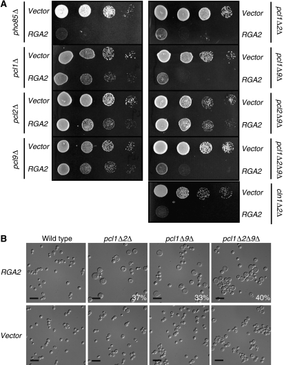
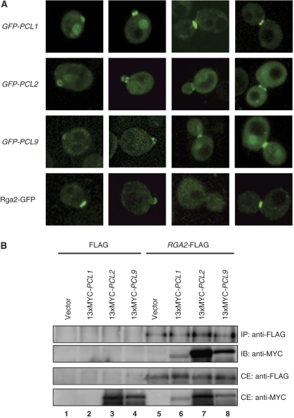
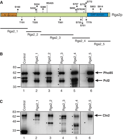
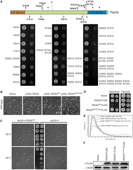
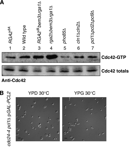
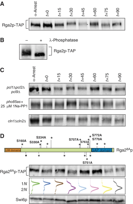
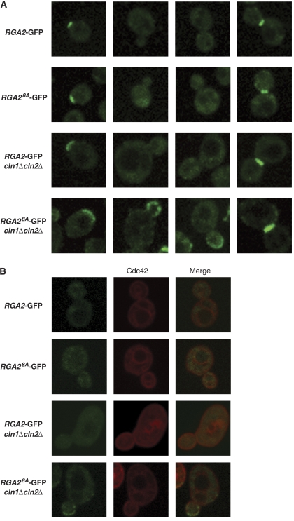
Cyclin-dependent kinases (CDKs) trigger essential cell cycle processes including critical events in G1 phase that culminate in bud emergence, spindle pole body duplication, and DNA replication. Localized activation of the Rho-type GTPase Cdc42p is crucial for establishment of cell polarity during G1, but CDK targets that link the Cdc42p module with cell growth and cell cycle commitment have remained largely elusive. Here, we identify the GTPase-activating protein (GAP) Rga2p as an important substrate related to the cell polarity function of G1 CDKs. Overexpression of RGA2 in the absence of functional Pho85p or Cdc28p CDK complexes is toxic, due to an inability to polarize growth. Mutation of CDK consensus sites in Rga2p that are phosphorylated both in vivo and in vitro by Pho85p and Cdc28p CDKs results in a loss of G1 phase-specific phosphorylation. A failure to phosphorylate Rga2p leads to defects in localization and impaired polarized growth, in a manner dependent on Rga2p GAP function. Taken together, our data suggest that CDK-dependent phosphorylation restrains Rga2p activity to ensure appropriate activation of Cdc42p during cell polarity establishment. Inhibition of GAPs by CDK phosphorylation may be a general mechanism to promote proper G1-phase progression.


Figures

Figure 1
Figure 1
G1 cyclin mutants are sensitive to overexpression of RGA2. (A) Growth defect caused by overexpression of RGA2 in the absence of PHO85, PHO85 G1 cyclins, or CDC28 G1 cyclins. Isogenic wild-type, pho85Δ, pcl1Δ, pcl2Δ, pcl9Δ, pcl1Δpcl2Δ, pcl2Δpcl9Δ, pcl1Δpcl9Δ, pcl1Δpcl2Δpcl9Δ, and cln1Δcln2Δ strains bearing either pGST-RGA2 or vector were spotted in serial 15-fold dilutions on medium containing galactose and incubated at 30°C for 72 h. (B) Overexpression of RGA2 in G1 phase-specific Pho85p cyclin mutants results in an accumulation of large, round, and unbudded cells. The morphology of those strains (top panels) from panel A was examined following 72 h of growth on galactose-containing medium. Cells were visualized at × 630 magnification. Size bar is 10 μm. The number in the bottom right corner refers to the percentage of cells displaying a large, unbudded phenotype within the population relative to the vector control (bottom panel); >200 cells were counted for each sample.
Figure 2
Figure 2
G1-specific Pho85 cyclins have overlapping localization and interact physically with Rga2p. (A) Localization of G1-specific Pho85 cyclins to sites of polarized growth. The localization of GFP-tagged Pcls was examined by confocal microscopy following expression of GFP-PCL1, GFP-PCL2, or GFP-PCL9 from their native promoters on high-copy plasmids. The localization pattern of Rga2p-GFP expressed from its native chromosomal locus is also shown (bottom). (B) Co-immunoprecipitation of G1-specific Pcl cyclins with Rga2p. Extracts from strains coexpressing FLAG and vector, or RGA2-FLAG and vector (lanes 1 and 5, respectively); FLAG and 13xMYC-PCL1, or RGA2-FLAG and 13xMYC-PCL1 (lanes 2 and 6, respectively); FLAG and 13xMYC-PCL2, or RGA2-FLAG and 13xMYC-PCL2 (lanes 3 and 7, respectively); FLAG and 13xMYC-PCL9, or RGA2-FLAG and PCL9-13xMYC (lanes 4 and 8, respectively), were used to immunoprecipitate (IP) Rga2p. Rga2p-FLAG was detected by Western blot analysis using anti-FLAG antibodies. 13xMYC-tagged Pcls were detected by immunoblotting (IB) with anti-MYC antibodies. A total of 10% of input cell extract was loaded as a control (CE).
Figure 3
Figure 3
G1-specific CDKs can phosphorylate Rga2 in vitro. (A) Schematic representation of full-length Rga2p. Locations of potential CDK phosphorylation sites (S/TP) are indicated by asterisks. The regions of Rga2p contained in five fragments used in kinase assays are shown (Rga2_1 through Rga2_5). (B) Phosphorylation of Rga2p by Pcl2p-Pho85p kinase in vitro. The five GST-Rga2p fragments (see A) were mixed with Pcl2p–Pho85p (lanes 2–6) in kinase reactions along with [γ-32P]ATP. Pho4p (lane 1) was included as a control. Phosphorylation of proteins was analyzed by SDS–PAGE and autoradiography. The position of migration of input proteins (see Supplementary Figure 2 for Coomassie Blue-stained gel) is indicated by stars. The positions of migration of phosphorylated Pcl2p and auto-phosphorylated Pho85p are indicated. (C) Phosphorylation of Rga2p by Cln2p–Cdc28p kinase in vitro. GST-Rga2p fragments were mixed with Cln2p–Cdc28p kinase (lanes 2–6) in kinase reactions. Histone H1 (lane 1) was included as a control. The position of migration of input proteins (see Supplementary Figure 2 for Coomassie Blue-stained gel) is indicated by stars. The position of phosphorylated Cln2p is indicated.
Figure 4
Figure 4
Functional analysis of RGA2 mutations. (A) A wild-type strain bearing either vector, pGAL-RGA2, or plasmids expressing RGA2 mutated at potential CDK phosphorylation sites was spotted in serial 15-fold dilutions on galactose medium and incubated at 30°C for 72 h. The schematic of full-length Rga2p shows those potential CDK phosphorylation sites (asterisks; S/TP) mutated to alanine. The version comprising the eight alanine substitutions, S330A, S334A, S707A, S751A, S160A, S763A, S770A, S772A, is referred to as RGA28A. (B) Wild type strains carrying either vector, pGAL-RGA2-FLAG, pGAL-RGA28A-FLAG, or pGAL-RGA28A,K872A-FLAG were induced in galactose for 6 h and examined by microscopy. Cells were visualized at × 400 magnification. Size bar is 10 μm. The number in the bottom right corner refers to the percentage of cells displaying a large, unbudded phenotype relative to the vector control; >400 cells were counted. (C) A cdc24-4 strain bearing the RGA28A allele at the endogenous RGA2 locus was spotted in serial 15-fold dilutions on rich medium, and incubated at semi-permissive temperatures. Cell morphology was examined using differential interference contrast (DIC) microscopy at × 400 magnification. Size bar is 10 μm. (D) Wild-type strains bearing either vector, pGAL-RGA2, pGAL-RGA28A, or pGAL-RGA28A,K872A were spotted in serial 15-fold dilutions on galactose- (left) or glucose (right)-containing medium and incubated at 30°C for 72 h. (E) Wild-type cells bearing either vector, pGAL-RGA2, pGAL-RGA28A, or pGAL-RGA28A,K872A were grown to log phase and induced with galactose for 6 h. The cell volume distribution (fl) in culture was measured using a Coulter Z2 Particle analyzer. The median volume of cells was as follows: vector=36±5 fl; RGA2=40±1 fl; RGA28A=67±1 fl; and RGA28A,K872A=42±1 fl. (F) RGA2-FLAG, RGA28A-FLAG, and RGA28A,K872A-FLAG were expressed in low copy under the regulation of the GAL1 promoter in wild-type cells. Cells were induced with galactose for 3 h. Expression was monitored by Western blot analysis with α-FLAG antibodies (top panel) and compared to levels of Swi6 detected in extracts using α-Swi6 antibodies (bottom panel).
Figure 5
Figure 5
Levels of activated Cdc42p are reduced in CDK mutants and in wild-type cells expressing RGA28A. (A) Cell extract from a strain bearing RGA28A at the RGA2 endogenous locus (lane 1), a wild-type strain (lane 2), an RGA28Abem3Δrga1Δ strain (lane 3), an rga2Δbem3Δrga1Δ triple GAP mutant (lane 4), a pho85Δ mutant (lane 5), a clnΔcln2Δmutant (lane 6), and a pcl1Δ2Δ9Δmutant (lane7) were incubated with GST-PAK (CRIB) beads that bind Cdc42-GTP. Cdc42p was detected by Western blot using α-Cdc42 antibodies. (B) Removal of the Pho85 G1 cyclins, PCL1 and PCL2, exacerbates the phenotype of cdc24-4 allele-bearing cells. A cdc24-4 pcl1ΔpGAL-3xHA-PCL2 strain was grown in the presence of glucose (YPD) or galactose (YPG), at a semi-permissive temperature of 30°C, and examined by microscopy. Cells were visualized at × 400 magnification. Size bar is 10 μm.
Figure 6
Figure 6
Rga28Ap fails to accumulate G1 phase-specific phosphoforms. (A) The phosphorylation of Rga2p-TAP during a cell cycle was monitored by Western blotting using α-TAP antibody. Samples were taken every 15 min for 90 min following alpha-factor block and release. (B) Rga2p-TAP was immunoprecipitated, divided into two aliquots, and one aliquot was treated with 100 U of lambda phosphatase. (C) The phosphorylation of Rga2p-TAP in pcl1Δpcl2Δpcl9Δ, pho85-as, and cln1Δcln2Δ cells was monitored during a cell cycle by Western blotting using polyclonal α-TAP antibodies. Samples were taken every 15 min for 90 min following alpha-factor block and release. For the pho85-as (analogue sensitive) strain, cells were released from alpha-factor into the indicated concentration of 1Na-PP1. (D) The phosphorylation of Rga2p-TAP or Rga28Ap-TAP in wild-type cells was monitored during a cell cycle by Western blotting using α-TAP antibody. Samples were taken every 15 min for 90 min following alpha-factor block and release. Corresponding FACS profiles indicate relative position in the cell cycle. Western blot analysis of Swi6p was used to assess loading. The schematic of Rga28Ap shows those potential CDK phosphorylation sites (asterisks; S/TP) mutated to alanine.
Figure 7
Figure 7
Localization of Rga2p in late G1 phase is dependent on phosphorylation by Pho85p and Cdc28p CDK complexes. (A) Phosphorylation influences the localization of Rga2p in cln1Δcln2Δ cells. Wild-type cells expressing RGA2-GFP or RGA28A-GFP, and cln1Δcln2Δ cells expressing RGA2-GFP or RGA28A-GFP were examined using spinning-disc confocal microscopy. Individual cells representing various stages of the cell cycle are highlighted. (B) RGA28A shares overlapping localization with Cdc42p in cln1Δcln2Δ cells. Those strains from panel A were transformed with pMET-mcherry-CDC42 and examined using spinning-disc confocal microscopy.

Similar articles

Cited by

References

    1. Aguilar RC, Longhi SA, Shaw JD, Yeh LY, Kim S, Schon A, Freire E, Hsu A, McCormick WK, Watson HA, Wendland B (2006) Epsin N-terminal homology domains perform an essential function regulating Cdc42 through binding Cdc42 GTPase-activating proteins. Proc Natl Acad Sci USA 103: 4116–4121 - PMC - PubMed
    1. Archambault V, Chang EJ, Drapkin BJ, Cross FR, Chait BT, Rout MP (2004) Targeted proteomic study of the cyclin–Cdk module. Mol Cell 14: 699–711 - PubMed
    1. Benton BK, Tinkelenberg AH, Jean D, Plump SD, Cross FR (1993) Genetic analysis of Cln/Cdc28 regulation of cell morphogenesis in budding yeast. EMBO J 12: 5267–5275 - PMC - PubMed
    1. Bishop AC, Ubersax JA, Petsch DT, Matheos DP, Gray NS, Blethrow J, Shimizu E, Tsien JZ, Schultz PG, Rose MD, Wood JL, Morgan DO, Shokat KM (2000) A chemical switch for inhibitor-sensitive alleles of any protein kinase. Nature 407: 395–401 - PubMed
    1. Bloom J, Cross FR (2007) Multiple levels of cyclin specificity in cell-cycle control. Nat Rev Mol Cell Biol 8: 149–160 - PubMed

Publication types

Substances