Review
. 2007 Oct;7(10):778-89.
doi: 10.1038/nri2172.

Calcium signalling and cell-fate choice in B cells

Affiliations
Review

Calcium signalling and cell-fate choice in B cells

Andrew M Scharenberg et al. Nat Rev Immunol. 2007 Oct.

Abstract

Alterations in the cytosolic concentration of calcium ions (Ca2+) transmit information that is crucial for the development and function of B cells. Cytosolic Ca2+ concentration is determined by a balance of active transport and gradient-driven Ca2+ fluxes, both of which are subject to the influence of multiple receptors and environmental sensing pathways. Recent advances in genomics have allowed for the compilation of an increasingly comprehensive list of Ca2+ transporters and channels expressed by B cells. The increasing understanding of the function and regulation of these proteins has begun to shift the frontier of Ca2+ physiology in B cells from molecular analysis to determining how diverse inputs to cytosolic Ca2+ concentration are integrated in specific immunological contexts.


Figures

Figure 1
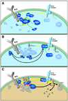
Figure 1. Ca2+ physiology in B cells at rest and during InsP3-mediated Ca2+ signal
A: In "resting" B cells, cytoplasmic Ca2+ homeostasis is maintained primarily through the actions of plasma membrane Ca2+ ATPase (PMCA) and sarcoplasmic/endoplasmic reticulum Ca2+ ATPase (SERCA) active transporters or pumps. Na+/Ca2+ exchange is limited due to lack of cytoplasmic Ca2+, and plasma membrane channels are primarily in the closed state. B: A receptor-mediated (inositol-1,4,5-trisphosphate (IP3))-dependent calcium signal is initiated through binding of IP3 to IP3R proteins in the endoplasmic reticulum (ER) membrane. Opening of IP3R channels allows Ca2+ to enter the cytoplasm from ER Ca2+ stores. Once ER Ca2+ stores are sufficiently depleted, ER Stromal interaction molecule (STIM) family proteins are activated to move into proximity to, and open, Ca2+-release activated channels (CRAC channels) in plasma membrane microdomains. Notable aspects of cell physiology in the context of an active Ca2+ signal include: (1) plasma membrane potential provides a significant driving force for Ca2+ entry through ion channels; (2) active Ca2+ transport activities (sarcoplasmic reticulum/endoplasmic reticulum Ca2+ ATPase (SERCA), plasma membrane Ca2+ ATPase (PMCA), and Na+/Ca2+ exchange) increase relative to the resting state in response to the increased availability of Ca2+ on their pumping cytosolic surfaces; and (3) Na+ entry into the cytoplasm may significantly influence cytosolic Ca2+ concentration depending on local changes in Na+ concentrations and the relative flow of Ca2+ through Ca2+ selective ion channels, non-selective cation channels (G), and reverse mode Na+ /Ca2+ exchange (that is, Ca2+ transported to the cytosol from the extracellular environment in exchange for intracellular Na+)–.
Figure 2
Figure 2. Biochemical events in a BCR signaling complex leading to amplified PLCγ2 activation
A: Recruitment and phosphorylation (yellow arrows and dots) of B-cell linker (BLNK) protein within the signalosome of the B-cell receptor (BCR) generates binding sites and recruitment (green arrows) of key BCR effectors VAV, GRB2 (growth-factor-receptor-bound protein 2), phospholipase Cg2 (PLCγ2) and Bruton's tyrosine kinase (BTK). B: P110 Phosphoinositide-3-kinase (PI3K) activation occurs through several parallel pathways (black arrows), and leads to local phosphorylation (yellow arrow) of phosphatidylinositol-4,5-bisphosphate to phosphatidylinositol-3,4,5-trisphosphate (PtdIns(3,4,5)P3). This effect is amplified in part through recruitment (green arrow) of the p85 subunit of PI3K to the CD19 complex. C: Recruitment of BTK to PtdIns(3,4,5)P3 induces its transphosphorylation by lyn (small yellow arrows), and initiates a positive feedback loop (large yellow arrows): association of BTK with phosphatidyl inositol-4-phosphate 5-kinase (PI5K) leads to increased production of phosphatidylinositol-4,-5- bisphosphate (PtdIns(4,5)P2) and so increased substrate for PtdIns(3,4,5)P3production; enhanced PtdIns(3,4,5)P3 leads to increased BTK recruitment (with additional PI5K) and BTK activation. The overall signal is read out through BTK-dependent tyrosine phosphorylation and activation of PLCγ2 (red arrows), which hydrolyzes (black arrows) locally available and newly synthesized PtdIns(4,5)P2 to IP3 and diacylglycerol (DAG).
Figure 3
Figure 3. Surface-receptor mechanisms for modulation of BCR-induced Ca2+ signals
The CD19 complex is a positive regulatory receptor. Its active recruitment of into B-cell receptor (BCR) signaling complexes acts to enhance p110 phosphoinositide-3-kinase (PI3K) activation during BCR signaling. FcγRIIb1 and CD22 are both negative regulators of BCR-mediated calcium signals. When actively recruited into BCR signaling complexes, these receptors both act by recruiting the SH2-containing tyrosine phosphatase-1 (SHP1) and the SH2-containing inositol-5'-phosphatase (SHIP). However, the majority of the impact of FcγRIIb1 on Ca2+ signaling is mediated through the SHIP-dependent attentuation of PI3K signaling, while CD22 appears to have its most significant effect on Ca2+ signaling through SHP-1-dependent enhancement of PMCA-mediated Ca2+ export.
Figure 4
Figure 4. Mechanisms for regulation of cytosolic Ca2+ in B cells
Pathways with known or potential ability to influence cytosolic Ca2+ in B-cells are grouped by colour according to their mechanism of influence.
Figure 5
Figure 5. Membrane potential modulation of Ca2+ signals
Membrane potential can be differentially and dynamically regulated through K+ channel activity and TRPM4/TRPM5 monovalent cation channel activity Top panel: In resting cells, membrane potential is set at a highly negative potential through the activity of voltage–operated K+ channels. The negative membrane potential is generated through the outward movement of positively charged K+ ions, leaving unpaired intracellular negative charges (not shown). TRPM4/TRPM5 channels are thought to be closed in resting cells. Middle panel: When a cell stimulus leads to activation of the store-operated Ca2+ entry (SOCE) pathway, opening of highly Ca2+-selective CRAC channels allows Ca2+ to pass across the plasma membrae. The current carried by CRAC channels is very small, and thus the entering positively charged Ca2+ ions have little direct influence on the membrane potential. However, the rise in cytosolic Ca2+ resulting from CRAC channel opening may influence the gating of both Ca2+-activated K+ channels and TRPM4/TRPM5 channels. Bottom left panel: In cells expressing significant numbers of Ca2+-activated K+ channels, their activation serves to maintain a highly negative membrane potential. Thus, the cell maintains a strong sustained driving force for Ca2+ entry and a large magnitude sustained Ca2+ signal results. Bottom right panel: In cells expressing significant numbers of TRPM4/TRPM5 monovalent cation channels, their activation allows Na+ entry, thus providing cytosolic positive ions to neutralize the unpaired internal charges. This diminishes the negative membrane potential, thereby attenuating the driving force for Ca2+ entry, and resulting in a reduced magnitude Ca2+ signal. Beyond Ca2+-dependent activation of K+ and TRPM4/TRPM5 channels, the activity of these channels is variously accessible to receptor mediated stimuli, metabolic mediators, and environmental influences that modulate their gating properties, providing multiple mechanisms through which dynamic regulation of membrane potential could influence B-cell Ca2+ signaling.
Figure 6
Figure 6. Potential mechanisms for Ca2+-dependent modulation of B-cell fate determination
Based on present models for Ca2+-dependent regulation of transcription factors such as Nuclear Factor of Activated T-cells (NFAT) and Nuclear Factor kb (NF-κB), membrane potential modulation and direct Ca2+ entry through non-SOCE pathways are predicted to have the capacity to significantly alter B-cell-fate choice. A: Strong B-cell receptor (BCR) activation fully activates both NF-κB (through inhibitor of kb (IκB) degradation) and NFAT (through calcineurin phosphatase activation) pathways. B: Modulation of membrane potential attenuates late Ca2+ entry, selectively influencing NFAT activation. C: A weak but sustained BCR activation signal activates only the NFAT pathway. D: Late activation of a direct Ca2+ entry pathway restores NF-κB pathway activation despite a weak BCR activation signal.

Similar articles

Cited by

References

    1. Quintana A, Griesemer D, Schwarz EC, Hoth M. Calcium-dependent activation of T-lymphocytes. Pflugers Arch. 2005;450:1–12. - PubMed
    1. Lewis RS. Calcium signaling mechanisms in T lymphocytes. Annu Rev Immunol. 2001;19:497–521. - PubMed
    1. Cahalan MD, Wulff H, Chandy KG. Molecular properties and physiological roles of ion channels in the immune system. J Clin Immunol. 2001;21:235–252. - PubMed
    1. Brose N, Betz A, Wegmeyer H. Divergent and convergent signaling by the diacylglycerol second messenger pathway in mammals. Curr Opin Neurobiol. 2004;14:328–340. - PubMed
    1. Rhee SG. Regulation of phosphoinositide-specific phospholipase C. Annu Rev Biochem. 2001;70:281–312. - PMC - PubMed

Publication types