

Enhancement of intratumoral cyclophosphamide pharmacokinetics and antitumor activity in a P450 2B11-based cancer gene therapy model
- PMID: 17853921
- PMCID: PMC2613860
- DOI: 10.1038/sj.cgt.7701092
Enhancement of intratumoral cyclophosphamide pharmacokinetics and antitumor activity in a P450 2B11-based cancer gene therapy model
Abstract
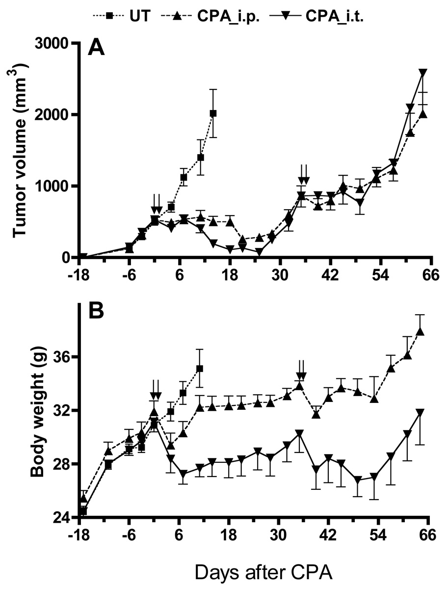
The therapeutic utility of cytochrome P450-based enzyme prodrug therapy is well established by preclinical studies and in initial clinical trials. The underlying premise of this gene therapy is that intratumoral P450 expression leads to in situ activation of anticancer P450 prodrugs, such as cyclophosphamide (CPA), with intratumoral accumulation of its activated 4-OH metabolite. In mice bearing 9L gliosarcomas expressing the CPA 4-hydroxylase P450 2B6, enhanced tumor apoptosis was observed 48 h after CPA treatment; however, intratumoral 4-OH-CPA levels were indistinguishable from those of P450-deficient tumors, indicating that the bulk of activated CPA is derived from hepatic metabolism. In contrast, in 9L tumors expressing P450 2B11, a low K(m) CPA 4-hydroxylase, intratumoral 4-OH-CPA levels were higher than in blood, liver and P450-deficient tumors. Intratumoral 4-OH-CPA increased dose-dependently, without saturation at 140 mg kg(-1) CPA, suggesting restricted tumor cell permeation of the parent drug. To circumvent this problem, CPA was administered by direct intratumoral injection, which increased the maximum concentration and area under the curve of drug concentration x time (AUC) of intratumoral 4-OH-CPA by 1.8- and 2.7-fold, respectively. An overall 3.9-fold increase in intratumoral 4-OH-CPA AUC, and in antitumor activity, was obtained when CPA release to systemic circulation was delayed using the slow-release polymer poloxamer 407 as vehicle for intratumoral CPA delivery. These findings highlight the advantage of gene therapy strategies that combine low K(m) P450 prodrug activation enzymes with slow, localized release of P450 prodrug substrates.
Figures


Similar articles
-
Impact of liver P450 reductase suppression on cyclophosphamide activation, pharmacokinetics and antitumoral activity in a cytochrome P450-based cancer gene therapy model.Cancer Gene Ther. 2000 Jul;7(7):1034-42. doi: 10.1038/sj.cgt.7700200. Cancer Gene Ther. 2000. PMID: 10917206
-
Enhanced antitumor activity of P450 prodrug-based gene therapy using the low Km cyclophosphamide 4-hydroxylase P450 2B11.Mol Cancer Ther. 2006 Mar;5(3):541-55. doi: 10.1158/1535-7163.MCT-05-0321. Mol Cancer Ther. 2006. PMID: 16546968
-
Collaboration between hepatic and intratumoral prodrug activation in a P450 prodrug-activation gene therapy model for cancer treatment.Mol Cancer Ther. 2007 Nov;6(11):2879-90. doi: 10.1158/1535-7163.MCT-07-0297. Epub 2007 Nov 7. Mol Cancer Ther. 2007. PMID: 17989319 Free PMC article.
-
Activation of oxazaphosphorines by cytochrome P450: application to gene-directed enzyme prodrug therapy for cancer.Toxicol In Vitro. 2006 Mar;20(2):176-86. doi: 10.1016/j.tiv.2005.06.046. Epub 2005 Nov 15. Toxicol In Vitro. 2006. PMID: 16293390 Review.
-
Cytochrome P450 gene-directed enzyme prodrug therapy (GDEPT) for cancer.Curr Pharm Des. 2002;8(15):1405-16. doi: 10.2174/1381612023394566. Curr Pharm Des. 2002. PMID: 12052216 Review.
Cited by
-
Modulation of the antitumor activity of metronomic cyclophosphamide by the angiogenesis inhibitor axitinib.Mol Cancer Ther. 2008 Jan;7(1):79-89. doi: 10.1158/1535-7163.MCT-07-0584. Mol Cancer Ther. 2008. PMID: 18202011 Free PMC article.
-
Dominant effect of antiangiogenesis in combination therapy involving cyclophosphamide and axitinib.Clin Cancer Res. 2009 Jan 15;15(2):578-88. doi: 10.1158/1078-0432.CCR-08-1174. Clin Cancer Res. 2009. PMID: 19147763 Free PMC article.
-
A multipurpose brachytherapy catheter to enable intratumoral injection.Brachytherapy. 2021 Jul-Aug;20(4):900-910. doi: 10.1016/j.brachy.2020.10.012. Epub 2021 Mar 27. Brachytherapy. 2021. PMID: 33785280 Free PMC article.
-
Advances in image-guided intratumoral drug delivery techniques.Ther Deliv. 2010 Aug;1(2):307-22. doi: 10.4155/tde.10.20. Ther Deliv. 2010. PMID: 22816134 Free PMC article. Review.
-
Oxazaphosphorine bioactivation and detoxification The role of xenobiotic receptors.Acta Pharm Sin B. 2012 Apr 1;2(2):10.1016/j.apsb.2012.02.004. doi: 10.1016/j.apsb.2012.02.004. Acta Pharm Sin B. 2012. PMID: 24349963 Free PMC article.
References
-
- McKeown SR, Ward C, Robson T. Gene-directed enzyme prodrug therapy: a current assessment. Curr Opin Mol Ther. 2004 Aug;6(4):421–435. - PubMed
-
- Shinohara ET, Lu B, Hallahan DE. The use of gene therapy in cancer research and treatment. Technol Cancer Res Treat. 2004 Oct;3(5):479–490. - PubMed
-
- Dachs GU, Tupper J, Tozer GM. From bench to bedside for gene-directed enzyme prodrug therapy of cancer. Anticancer Drugs. 2005 Apr;16(4):349–359. - PubMed
-
- Roy P, Waxman DJ. Activation of oxazaphosphorines by cytochrome P450: application to gene-directed enzyme prodrug therapy for cancer. Toxicol In Vitro. 2006 Mar;20(2):176–186. - PubMed
-
- Jounaidi Y. Cytochrome P450-based Gene Therapy for Cancer Treatment: From Concept to the Clinic. Curr Drug Metab. 2002 Dec;3(6):609–622. - PubMed
Publication types
MeSH terms
Substances
Grants and funding
LinkOut - more resources
Full Text Sources
Other Literature Sources
Medical
