. 2007 Oct;117(10):3097-106.
doi: 10.1172/JCI30602.

TREM-1--expressing intestinal macrophages crucially amplify chronic inflammation in experimental colitis and inflammatory bowel diseases

Affiliations

TREM-1--expressing intestinal macrophages crucially amplify chronic inflammation in experimental colitis and inflammatory bowel diseases

Mirjam Schenk et al. J Clin Invest. 2007 Oct.

Abstract

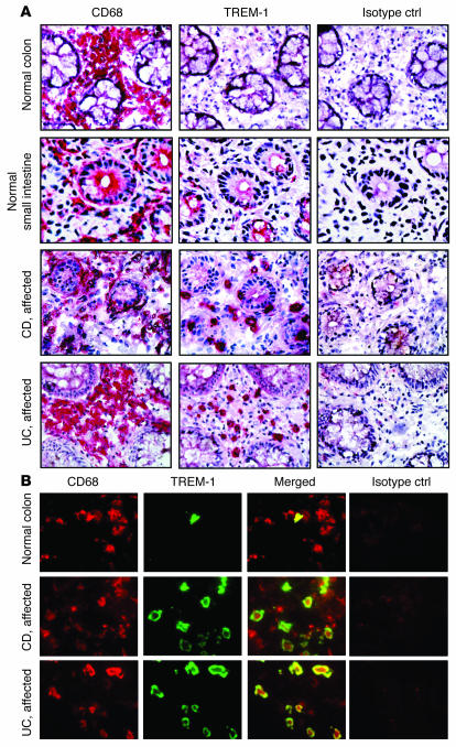
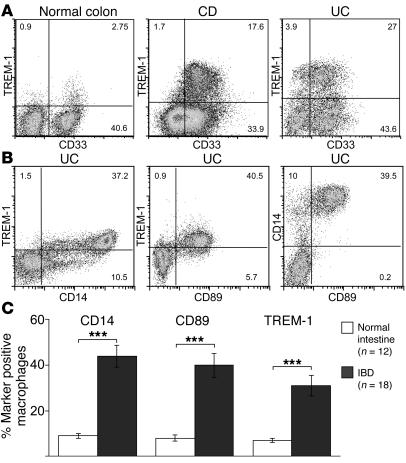
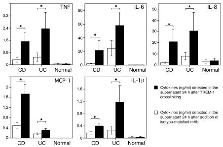
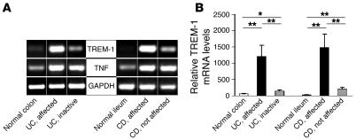
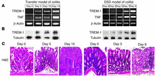
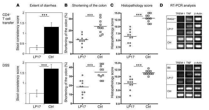
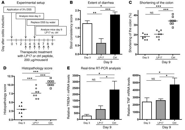
Triggering receptor expressed on myeloid cells-1 (TREM-1) potently amplifies acute inflammatory responses by enhancing degranulation and secretion of proinflammatory mediators. Here we demonstrate that TREM-1 is also crucially involved in chronic inflammatory bowel diseases (IBD). Myeloid cells of the normal intestine generally lack TREM-1 expression. In experimental mouse models of colitis and in patients with IBD, however, TREM-1 expression in the intestine was upregulated and correlated with disease activity. TREM-1 significantly enhanced the secretion of relevant proinflammatory mediators in intestinal macrophages from IBD patients. Blocking TREM-1 by the administration of an antagonistic peptide substantially attenuated clinical course and histopathological alterations in experimental mouse models of colitis. This effect was also seen when the antagonistic peptide was administered only after the first appearance of clinical signs of colitis. Hence, TREM-1-mediated amplification of inflammation contributes not only to the exacerbation of acute inflammatory disorders but also to the perpetuation of chronic inflammatory disorders. Furthermore, interfering with TREM-1 engagement leads to the simultaneous reduction of production and secretion of a variety of pro-inflammatory mediators such as TNF, IL-6, IL-8 (CXCL8), MCP-1 (CCL2), and IL-1beta. Therefore, TREM-1 may also represent an attractive target for the treatment of chronic inflammatory disorders.


Figures

Figure 1
Figure 1. TREM-1–expressing macrophages are increased in the intestinal lamina propria of IBD patients.
(A) Serial frozen sections of the intestine from normal donors and IBD (CD and UC) patients with active disease were immunostained for TREM-1 and CD68. An isotype-matched irrelevant mAb was used as a negative control (Isotype ctrl). Stainings are representative of at least 12 different intestinal tissue samples for the normal small and large intestine and of at least 10 tissues each for UC and CD. Original magnification, ×40. (B) Double immunofluorescence stainings demonstrate that most TREM-1–positive cells (green) in the intestinal lamina propria from patients with active IBD (UC and CD) were CD68+ macrophages (red). Original magnification, ×100.
Figure 2
Figure 2. TREM-1–expressing macrophages are increased in the affected intestinal mucosa of patients with IBD and coexpress CD14 and CD89.
(A) Lamina propria macrophages of normal intestinal tissue specimens and of patients with active IBD were isolated and analyzed by FACS for TREM-1, CD14, and CD89 cell-surface expression. (B) Coexpression of CD14 and CD89 on TREM-1–positive intestinal macrophages is shown for a representative UC patient. (C) The frequency of TREM-1–expressing lamina propria macrophages is significantly increased in patients with IBD (n = 18: UC, n = 7; CD, n = 11) compared with normal individuals (n = 12). Percentages are indicated as mean ± SEM; ***P < 0.001.
Figure 3
Figure 3. Secretion of TNF, IL-6, IL-8 (CXCL8), monocyte chemoattractant protein–1 (MCP-1 [CCL2]), and IL-1β is significantly enhanced following engagement of TREM-1 on intestinal lamina propria macrophages from IBD but not from normal control patients.
Intestinal macrophages isolated from CD patients (n = 11), UC patients (n = 7), and normal control patients (n = 5) were cultured for 24 hours in the presence (black bars) or absence (white bars) of an agonistic plate-bound anti–TREM-1 mAb. Cytokine concentrations were measured simultaneously in the supernatant. Data represent mean ± SEM; *P < 0.05.
Figure 4
Figure 4. Enhanced disease activity is associated with increased TREM-1 mRNA expression in the intestinal mucosa of patients with IBD.
RNA was isolated from endoscopic biopsies of actively inflamed intestinal mucosa and of macroscopically nonaffected areas from CD and UC patients and from normal control samples. (A) Semiquantitative RT-PCR was performed for TREM-1 and TNF. Amplification of GAPDH is shown as an internal control. One representative example is shown for 5 normal, 11 CD, and 7 UC patients analyzed. (B) TREM-1 mRNA levels were analyzed by quantitative RT-PCR. The cycle number at which the TREM-1 transcripts were detectable was compared with that of GAPDH as an internal control and expressed as arbitrary units; indicated are the mean values (± SEM) from at least 6 tissue samples per group. **P < 0.01; *P < 0.05.
Figure 5
Figure 5. TREM-1 mRNA and protein expression in the colon correlates with colonic inflammation and is induced early during onset of experimental colitis.
(A) Kinetics of TREM-1 and TNF transcription in the colon upon colitis induction by CD4+ T cell transfer into RAG2–/– recipient mice (transfer model) and the DSS mouse model of intestinal inflammation in C57BL/6 mice. β-Actin was used as a loading control. (B) Western blot analyses of colonic tissue samples adjacent to those used for RNA isolation demonstrate the increased TREM-1 protein expression during induction of colitis. Tubulin is shown as a control. (C) H&E-stained paraffin-embedded tissue sections demonstrate the onset of histopathological alterations after 5 days after cell transfer and after 3 days after DSS administration. Original magnification, ×40. In each model 4 mice were analyzed per time point in 2 independent experiments.
Figure 6
Figure 6. Blocking TREM-1 attenuates intestinal inflammation in 2 distinct mouse models of colitis.
Colitis was induced either by the transfer of splenic CD4+CD45RBhi T cells into RAG2–/– recipients (top row) or by oral administration of DSS (bottom row). Upon colitis induction, experimental mice were treated daily either with an antagonistic TREM-1 peptide (LP17) or with a control peptide. (A) Fecal samples from each mouse were tested for the presence of occult blood and bloody diarrhea to obtain the stool consistency score (see Methods). Data are presented as mean ± SEM; n = 10. (B) The colon length of each mouse was measured from the end of the cecum to the anus; indicated are the mean loss of colon length (%) at the end of the experiment. (C) The colitis scores for the 2 groups (LP17-treated and control-treated) were determined as described in Methods. (D) PCR analysis for TREM-1 and TNF mRNA was performed on identical segments of the distal colon in untreated mice (RAG2–/–; C57BL/6 [B6]) and antagonistic TREM-1– (LP17-) and control peptide–treated mice upon colitis induction. β-Actin was used as loading control. Results for 1 representative unmanipulated control mouse and 2 representative experimental mice per group are shown. ***P < 0.001; n = 10.
Figure 7
Figure 7. Blocking TREM-1 attenuates the disease process in established colitis.
(A) When after oral administration of DSS the experimental mice developed colitis with persistent diarrhea and fecal samples had tested positive for the presence of occult blood (day 3), the first group of mice was analyzed (day 3; n = 8), and the remaining experimental mice were treated daily with either antagonistic TREM-1 peptide (LP17; n = 8) or with control peptide (n = 8) for an additional 5 days. (B) Fecal samples from each mouse were tested for the presence of occult blood and bloody diarrhea to obtain the stool consistency score (see Methods). Data are indicated as mean ± SEM; n = 8. (C) The colonic length in each mouse was measured from the end of the cecum to the anus; indicated are the mean loss of colon length (%) on day 3 and on day 9 for both the LP17-treated and control-treated groups. (D) Colitis scores for the 3 groups of mice were determined as described in Methods. (E) Real-time RT-PCR analyses for TREM-1 and TNF mRNA were performed individually on identical segments of the distal colon from each mouse in all experimental groups. ***P < 0.001; **P < 0.01; *P < 0.05.

References

    1. Bouchon A., Cella M., Grierson H.L., Cohen J.I., Colonna M. Activation of NK cell-mediated cytotoxicity by a SAP-independent receptor of the CD2 family. J. Immunol. 2001;167:5517–5521. - PubMed
    1. Bouchon A., Facchetti F., Weigand M.A., Colonna M. TREM-1 amplifies inflammation and is a crucial mediator of septic shock. Nature. 2001;410:1103–1107. - PubMed
    1. Schenk M., Bouchon A., Birrer S., Colonna M., Mueller C. Macrophages expressing triggering receptor expressed on myeloid cells-1 are underrepresented in the human intestine. J. Immunol. 2005;174:517–524. - PubMed
    1. Bouchon A., Dietrich J., Colonna M. Cutting edge: inflammatory responses can be triggered by TREM-1, a novel receptor expressed on neutrophils and monocytes. J. Immunol. 2000;164:4991–4995. - PubMed
    1. Gibot S., et al. A soluble form of the triggering receptor expressed on myeloid cells-1 modulates the inflammatory response in murine sepsis. J. Exp. Med. 2004;200:1419–1426. - PMC - PubMed

Publication types

MeSH terms