

Liver angiogenesis as a risk factor for hepatocellular carcinoma development in hepatitis C virus cirrhotic patients
- PMID: 17854145
- PMCID: PMC4434626
- DOI: 10.3748/wjg.v13.i37.5009
Liver angiogenesis as a risk factor for hepatocellular carcinoma development in hepatitis C virus cirrhotic patients
Abstract
Aim: To evaluate the predictive value of hepatocyte proliferation and hepatic angiogenesis for the occurrence of Hepatocellular carcinoma (HCC) in hepatitis C virus (HCV) cirrhotic patients.
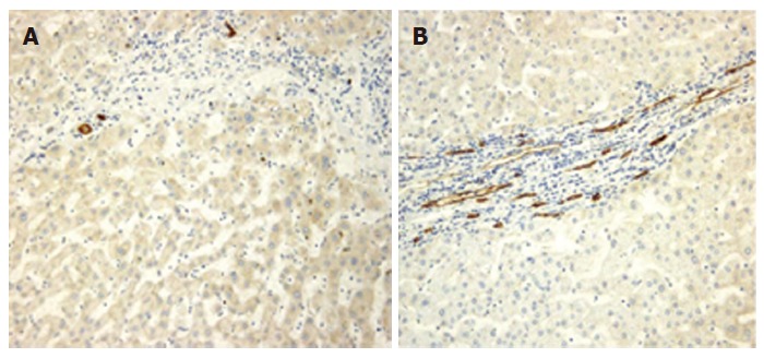
Methods: One hundred-five patients (69 males, 36 females; age range, 51-90 year; median 66 year) with biopsy proven HCV cirrhosis were prospectively monitored for HCC occurrence for a median time of 64 mo. Angiogenesis was assessed by using microvessel density (MVD), hepatocyte turnover by MIB1 and PCNA indexes at inclusion in liver biopsies.
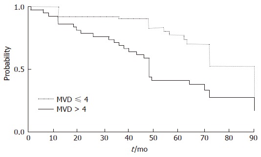
Results: Forty six patients (43.8%) developed HCC after a median time of 55 (6-120) mo while 59 (56.2%) did not. Patients were divided into two groups according to the median value of each index. The difference between patients with low (median MVD = 3; range 0-20) and high (median MVD = 7; range 1-24) MVD was statistically significant (chi(2) = 22.06; P < 0.0001) which was not the case for MIB1 or PCNA (MIB-1: chi(2) = 1.41; P = 0.2351; PCNA: chi(2) = 1.27; P = 0.2589). The median MVD was higher in patients who developed HCC than in those who did not. HCC-free interval was significantly longer in patients with the MVD < or = 4 (P = 0.0006). No relationship was found between MIB1 or PCNA and MVD (MIB-1 r(2) = 0.00007116, P = 0.9281; PCNA: r(2) = 0.001950; P = 0.6692). MVD only was able to predict the occurrence of HCC in these patients. Among other known risk factors for HCC, only male sex was statistically associated with an increased risk.
Conclusion: Liver angiogenesis has a role for in HCV-related liver carcinogenesis and for defining patients at higher risk.
Figures
References
-
- El-Serag HB. Hepatocellular carcinoma: an epidemiologic view. J Clin Gastroenterol. 2002;35:S72–S78. - PubMed
-
- Di Bisceglie AM. Epidemiology and clinical presentation of hepatocellular carcinoma. J Vasc Interv Radiol. 2002;13:S169–S171. - PubMed
-
- Block TM, Mehta AS, Fimmel CJ, Jordan R. Molecular viral oncology of hepatocellular carcinoma. Oncogene. 2003;22:5093–5107. - PubMed
-
- Romeo R, Colombo M. The natural history of hepatocellular carcinoma. Toxicology. 2002;181-182:39–42. - PubMed
-
- Hayashi J, Aoki H, Arakawa Y, Hino O. Hepatitis C virus and hepatocarcinogenesis. Intervirology. 1999;42:205–210. - PubMed
Publication types
MeSH terms
LinkOut - more resources
Full Text Sources
Other Literature Sources
Medical
Miscellaneous
