. 2007 Oct;66(2):468-83.
doi: 10.1111/j.1365-2958.2007.05935.x. Epub 2007 Sep 14.

The role of iron in Mycobacterium smegmatis biofilm formation: the exochelin siderophore is essential in limiting iron conditions for biofilm formation but not for planktonic growth

Affiliations

The role of iron in Mycobacterium smegmatis biofilm formation: the exochelin siderophore is essential in limiting iron conditions for biofilm formation but not for planktonic growth

Anil Ojha et al. Mol Microbiol. 2007 Oct.

Abstract

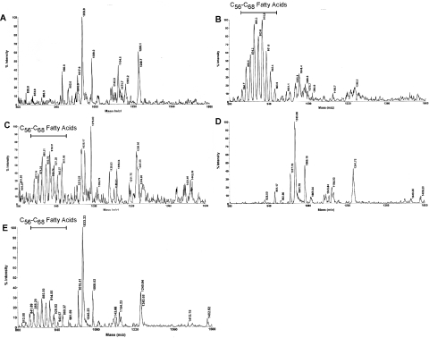
Many species of mycobacteria form structured biofilm communities at liquid-air interfaces and on solid surfaces. Full development of Mycobacterium smegmatis biofilms requires addition of supplemental iron above 1 microM ferrous sulphate, although addition of iron is not needed for planktonic growth. Microarray analysis of the M. smegmatis transcriptome shows that iron-responsive genes - especially those involved in siderophore synthesis and iron uptake - are strongly induced during biofilm formation reflecting a response to iron deprivation, even when 2 microM iron is present. The acquisition of iron under these conditions is specifically dependent on the exochelin synthesis and uptake pathways, and the strong defect of an iron-exochelin uptake mutant suggests a regulatory role of iron in the transition to biofilm growth. In contrast, although the expression of mycobactin and iron ABC transport operons is highly upregulated during biofilm formation, mutants in these systems form normal biofilms in low-iron (2 microM) conditions. A close correlation between iron availability and matrix-associated fatty acids implies a possible metabolic role in the late stages of biofilm maturation, in addition to the early regulatory role. M. smegmatis surface motility is similarly dependent on iron availability, requiring both supplemental iron and the exochelin pathway to acquire it.


Figures

Fig. 1
Fig. 1
Biofilm and planktonically grown cells used for microarray analyses. A. Biofilm cultures of wild-type strain of M. smegmatis mc2155 were grown in a modified M63 medium, either with or without supplemental iron (2 μM FeSO4) as shown, for 3, 4 or 5 days. RNA samples for microarray analysis were harvested from the 3 and 4 day plates grown in iron-supplemented medium. B. Planktonic growth of wild-type M. smegmatis mc2155 in no iron (blue), 2 μM iron (purple) or 50 μM iron (red). C. Growth curves of M. smegmatis mc2155 in liquid medium showing the increase in cell density (OD600; purple) and viable colony counts (cfu ml−1; blue). Arrows indicate the points at which cells were harvested for the exponentially growing planktonic and stationary phase to prepare samples for microarray analysis.
Fig. 2
Fig. 2
Schematic representations of transcriptome responses in 3 day biofilm, 4 day biofilm and stationary-phase conditions. A. Conditions corresponding to the 3 day biofilm (green circle), 4 day biofilm (blue circle) and stationary phase (red circle) are shown, with the numbers of induced genes (> 4-fold) shown in black and the number of repressed genes (> 4-fold) shown in red. The numbers of induced and regulated genes common to two or more conditions are included at the intersections of the three circles. A complete list of microarray data for genes in each category is included in Table S2. B. Induction of genes involved in iron acquisition during biofilm and stationary-phase growth. Twenty-nine genes arranged in nine operons (Msmeg0011–0014, Msmeg0015, Msmeg0016–0018, Msmeg2133–2135, Msmeg4502–4509, Msmeg4510, Msmeg5028, Msmeg6024–6026 and Msmeg6214–6216) putatively involved in iron acquisition show substantial levels of induction after 4 days of biofilm development. Arrows indicate the operon arrangements. Average fluorescence units are shown for the exponential planktonic (blue bars), 3 day biofilm (red bars), 4 day biofilm (yellow bars) and stationary phase (green bars). Fluorescence values for the exponential planktonic sample are the average of 12 normalized slides, and for the other three experimental conditions, are the average from three independent experiments.
Fig. 3
Fig. 3
Ferric exochelin biosynthesis (Msmeg0015) and uptake genes (Msmeg0011–0014) are required for biofilm formation but not for planktonic growth. A. Mutants defective in exochelin biosynthesis (ΔMsmeg0015, fxbA), exochelin uptake (ΔMsmeg0011–0014, fxuABC) and mycobactin biosynthesis (ΔMsmeg4509, mbtB) were generated by recombineering (van Kessel and Hatfull, 2007) and tested for biofilm growth. Growth after 4 days of biofilm development is shown using standard media containing a 2 μM iron supplement. All strains, including the wild-type control (mc2155), carry the pJV53 recombineering plasmid. B. Suppression of the biofilm defect of the mutants ΔMsmeg0011–0014 and ΔMsmeg0015 by addition of 50 μM iron to the growth medium. C. CAS-agar assay to test the synthesis and secretion of siderophore by ΔMsmeg0011–0014, ΔMsmeg0015, ΔMsmeg4509 and mc2155: pJV53 was used as a control. The orange halo around the colony is indicative of siderophore-mediated chelation of iron from CAS–iron complexes. D. Planktonic growth of the mutants described in A, compared with the parental M. smegmatis mc2155 strain. Cultures were grown in biofilm medium containing a 2 μM iron supplement.
Fig. 4
Fig. 4
Regulation of genes involved in siderophore synthesis and uptake. A. Analysis of expression of exochelin biosynthesis, fxbA (Msmeg0015), exochelin uptake, fxuC (Msmeg0012) and mycobactin synthesis, mbtB (Msmeg4509) genes by real-time RT-PCR. Expression levels were measured in samples grown for 3 or 4 days under biofilm conditions, and in early stationary phase, and represented as the relative log2 change in expression level compared with early planktonic growth. Similar samples were tested under the same growth conditions but in the presence of either 0, 2 or 50 μM iron as shown. B. Organization of the exochelin synthesis and uptake genes. The positions of gene between Msmeg0007 and Msmeg0019 are shown, along with their gene designations; Msmeg0008 and Msmeg0010 encode tRNAile and tRNAala genes respectively. Note that the organization differs somewhat from that described previously by Yu et al. (1998) in that Msmeg0011 is clearly part of an operon with Msmeg0012 (fxuC), Msmeg0013 (fxuB) and Msmeg0014 (fxuA). Putative IdeR iron boxes are shown as grey boxes. The lower panel shows the intergenic sequence between Msmeg0010 and Msmeg0011 that contains the putative regulatory features. The iron box consensus recognized by the IdeR regulator is shown above the location of a putative IdeR binding site (bold) upstream of Msmeg0011. The correspondence of this putative site to the consensus sequence (13 of the 19 positions are conserved) is similar to that for the known IdeR binding site upstream of fxbA (Msmeg0015) (Dussurget et al., 1996), and the position relative to the gene start site and putative −10 and −35 promoter is very similar. Arrows indicate the location of an imperfect inverted repeat that is a potential binding site for a second regulatory protein.
Fig. 5
Fig. 5
Iron is required for sliding motility of M. smegmatis. Colonies of either the parent strain (mc2155) or the iron uptake mutant (ΔMsmeg0011–0014) were placed at the centre of a sliding agar plate containing either 2 μM iron supplement or no iron (as indicated) and incubated at 37°C. Plates were photographed after 7 days.
Fig. 6
Fig. 6
Requirements of supplemental iron for biofilm formation and synthesis of C56–C68 fatty acids. M. smegmatis biofilms were grown for 4 days, with supplemental ferric iron induced in the following concentrations: none (A), 0.5 μM (B), 1 μM (C), 2 μM (D) and 5 μM (E). Samples were harvested, mycolic acids extracted, and analysed by MALDI-TOF as shown in the right. The position of the series of C56–C68 fatty acids is indicated above.
Fig. 7
Fig. 7
MALDI-TOF analysis of mycolic acid extracts from M. smegmatis cells in different growth states. The origin and treatment of cell cultures is as follows. A. Exponential planktonic growth, total-cell extract. B. Four-day biofilm, total-cell extract. C. Stationary-phase culture, total-cell extract. D. Four-day biofilm, cell-associated extract. E. Four-day biofilm, solvent extractable extract.
Fig. 8
Fig. 8
Response of fatty acid metabolism genes during biofilm formation and stationary phase. A. Of the 315 genes putatively involved in fatty acid metabolism, only seven clearly show induced expression (> 4-fold) in either the 3 or 4 day biofilm sample. Average fluorescence values of the seven genes are shown in exponential planktonic growth (blue bars), 3 day biofilms (red bars), 4 day biofilms (yellow bars) and stationary-phase cultures (green bars). Fluorescence values for the exponential planktonic sample are the average of 12 normalized slides, and for the other three experimental conditions, are the average from three independent experiments. B. M. smegmatis contains 42 putative fadE genes, most of which are not expressed under any of the conditions tested. Average fluorescence values for these 42 genes in exponential planktonic growth (blue bars), 3 day biofilms (red bars), 4 day biofilms (yellow bars) and stationary phase (green bars) are shown. The average values for each condition were calculated as described in panel A.

Similar articles

Cited by

References

    1. Andrews SC, Robinson AK, Rodriguez-Quinones F. Bacterial iron homeostasis. FEMS Microbiol Rev. 2003;27:215–237. - PubMed
    1. Banin E, Vasil ML, Greenberg EP. Iron and Pseudomonas aeruginosa biofilm formation. Proc Natl Acad Sci USA. 2005;102:11076–11081. - PMC - PubMed
    1. Bardarov S, Bardarov S, Pavelka MS, Sambandamurthy V, Larsen M, Tufariello J, et al. Specialized transduction: an efficient method for generating marked and unmarked targeted gene disruptions in Mycobacterium tuberculosis, M. bovis BCG and M. smegmatis. Microbiology. 2002;148:3007–3017. - PubMed
    1. Beenken KE, Dunman PM, McAleese F, Macapagal D, Murphy E, Projan SJ, et al. Global gene expression in Staphylococcus aureus biofilms. J Bacteriol. 2004;186:4665–4684. - PMC - PubMed
    1. Beloin C, Ghigo JM. Finding gene-expression patterns in bacterial biofilms. Trends Microbiol. 2005;13:16–19. - PubMed

Publication types

MeSH terms