. 2007 Sep 13:7:164.
doi: 10.1186/1471-2148-7-164.

Molecular evolution of type VI intermediate filament proteins

Affiliations

Molecular evolution of type VI intermediate filament proteins

Dominique Guérette et al. BMC Evol Biol. .

Abstract

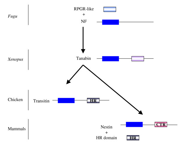
Background: Tanabin, transitin and nestin are type VI intermediate filament (IF) proteins that are developmentally regulated in frogs, birds and mammals, respectively. Tanabin is expressed in the growth cones of embryonic vertebrate neurons, whereas transitin and nestin are found in myogenic and neurogenic cells. Another type VI IF protein, synemin, is expressed in undifferentiated and mature muscle cells of birds and mammals. In addition to an IF-typical alpha-helical core domain, type VI IF proteins are characterized by a long C-terminal tail often containing distinct repeated motifs. The molecular evolution of type VI IF proteins remains poorly studied.

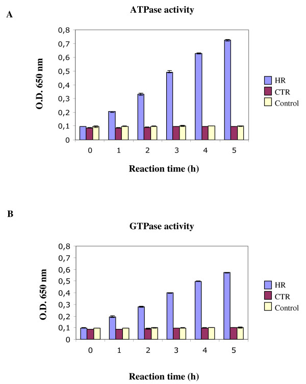
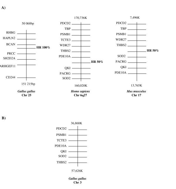
Results: To examine the evolutionary history of type VI IF proteins, sequence comparisons, BLAST searches, synteny studies and phylogenic analyses were performed. This study provides new evidence that tanabin, transitin and nestin are indeed orthologous type VI IF proteins. It demonstrates that tanabin, transitin and nestin genes share intron positions and sequence identities, have a similar chromosomal context and display closely related positions in phylogenic analyses. Despite this homology, fast evolution rates of their C-terminal extremity have caused the appearance of repeated motifs with distinct biological activities. In particular, our in silico and in vitro analyses of their tail domain have shown that (avian) transitin, but not (mammalian) nestin, contains a repeat domain displaying nucleotide hydrolysis activity.

Conclusion: These analyses of the evolutionary history of the IF proteins fit with a model in which type VI IFs form a branch distinct from NF proteins and are composed of two major proteins: synemin and nestin orthologs. Rapid evolution of the C-terminal extremity of nestin orthologs could be responsible for their divergent functions.


Figures

Figure 1
Figure 1
Schematic representation of the position of introns in type VI intermediate filament genes. A) The arrows along the tanabin cDNA diagram indicate the predicted positions of introns in the tanabin gene deduced from the genomic sequence of X. tropicalis (v4.1). B) The arrows indicate the location of introns in nestin [6], transitin [14] and synemin [13]. An IF protein diagram is illustrated at the bottom of the figure for orientation purposes. The blue box represents the α-helical rod domain.
Figure 2
Figure 2
Evolution of type VI IF proteins. A) Amino acid sequence alignment of the helix termination motif at the end of segment 2B of the α-helical rod domain of type VI IF proteins, desmin and NF-M from different vertebrate species. Asterisks represent amino acid residues specifically conserved in tanabin, transitin and nestin. The consensus sequence deduced from this multiple alignment is indicated at the bottom of the figure. The histogram indicates the level of residue conservation (from 0 to 100%) at each position. B) A phylogenetic tree was constructed based on the protein sequences of type IV NF-M and of type VI tanabin, transitin, nestin and synemin from different species using neighbor-joining methods (CLCbio software; 100 bootstraps). A closer evolutionary relationship is noticed between tanabin, transitin and nestin. The bootstrap values are shown at the nodes. xl, Xenopus laevis; xt, Xenopus tropicalis; gg, Gallus gallus; rn, Rattus norvegicus; mm, Mus musculus; hs, Homo sapiens.
Figure 3
Figure 3
Syntenic relationship among frog tanabin, avian transitin and nestin from human, mouse and rat. Physical maps of human, mouse, rat and chicken genomic sequences along with an anuran scaffold (X. tropicalis scaffold_790) were used to identify genes neighboring nestin, transtin and tanabin. Each diagram represents a chromosomal region whose position is defined by the number of base pairs from the telomere of the short arm of the chromosome (top and bottom of each diagram). Each horizontal line represents a gene position on the chromosome. Nestin, transitin and tanabin genes are shown in bold for clarity. Diagrams are arranged in such a way that the gene positions are visualized in the same orientation. LMNA, lamin A/C; RHGB, Rhesus blood group, B glycoprotein; HAPLN2, hyaluronan and proteoglycan linked protein 2; BCAN, brevican; PRCC, papillary renal cell carcinoma; SH2D2A, SH2 domain protein 2A; ARHGEF11, Rho guanine nucleotide exchange factor (GEF) 11 and CD244, CD244 natural killer cell receptor 2B4.
Figure 4
Figure 4
In vitro ATPase and GTPase activity of the HR domain. The HR domain of chicken transitin and the CTR domain of mouse nestin were expressed in bacteria and purified. Their ATPase/GTPase activities were tested individually using the malachite green colorimetric method for phosphate release determination. Phosphate release was determined at 25°C for each recombinant protein incubated with either A) ATP or B) GTP. The optical density at 650 nm was determined in triplicate at regularly timed intervals during the course of the enzymatic reaction and the mean values are presented with standard deviation error bars. Control: Buffer with the HR recombinant protein without NTP.
Figure 5
Figure 5
Reactivity of the HR-specific monoclonal antibody VAP-5 with the "hypothetical" mouse protein HRM. A fragment of mouse genomic clone RP2389A3 (gi: 106520665) showing the highest sequence identity to chick transitin HR domain has been subcloned in a pET-based expression vector and a His-tag-HRM fusion protein bacterially produced under IPTG induction. A co-migrating immunoreactive band was observed in Western blots using the anti-His-tag antibody and VAP-5 in extracts of bacteria submitted to IPTG induction but was not detected in the uninduced culture.
Figure 6
Figure 6
Syntenic relationships of the conserved HR domain in birds, humans and mice. Physical maps of human, mouse, rat and chicken genomic sequences were used to identify genes neighboring the conserved HR domains. Each diagram represents a chromosomal region whose position is defined by the number of base pairs from the telomere of the short arm of the chromosome (top and bottom of each diagram). Each horizontal line represents the position of a gene and is placed in physical order on the chromosome. Diagrams are depicted in such a way that genes are oriented in the same way. A) The HR domains were syntenic in human (chromosome 6q27) and mouse (chromosome 17). They share 50% identity with the chicken HR domain of transitin located on chromosome 25 (see Fig. 3). B) A corresponding gene cluster was found on chicken chromosome 3 from which an "avian" HR domain genomic segment was absent. RHGB, Rhesus blood group, B glycoprotein; HAPLN2, hyaluronan and proteoglycan link protein 2; BCAN, brevican; PRCC, papillary renal cell carcinoma; SH2D2A, SH2 domain protein 2A; ARHGEF11, Rho guanine nucleotide exchange factor (GEF) 11 and CD244, CD244 natural killer cell receptor 2B4; PDCD2, programmed cell death 2; PSMB1, proteasome (prosome, macropain) subunit, beta type, 1; TCTE3, t-complex-associated-testis-expressed 3; PDE10A, phosphodiesterase 10A; QKI, quaking homolog; SOD2, superoxide dismutase 2; THBS2, thrombospondin 2, PACRG, PARK2 co-regulated and WDR27, WD repeat domain 27.
Figure 7
Figure 7
Evolutionary model for type VI IF proteins. Fugu lacks obvious nestin homologs [38] and NF proteins may well represent ancestral type VI IF proteins [8]. Our model proposes that the first type VI IF protein resulted from the fusion of a RPGR-like cassette C-terminal to a NF gene. The C-terminal domain of a tanabin-like protein may have evolved giving rise to the HR domain displaying nucleotide hydrolysis activity in birds whereas this activity was progressively lost in mammalian nestins. In addition, the HR domain may have been duplicated and conserved in a mammalian gene that is not part of the IF family.

Similar articles

Cited by

References

    1. Hesse M, Magin TM, Weber K. Genes for intermediate filament proteins and the draft sequence of the human genome: novel keratin genes and a surprisingly high number of pseudogenes related to keratin genes 8 and 18. J Cell Sci. 2001;114:2569–2575. - PubMed
    1. Lazarides E. Intermediate filaments: a chemically heterogeneous, developmentally regulated class of proteins. Annu Rev Biochem. 1982;51:219–250. doi: 10.1146/annurev.bi.51.070182.001251. - DOI - PubMed
    1. Coulombe PA, Wong P. Cytoplasmic intermediate filaments revealed as dynamic and multipurpose scaffolds. Nat Cell Biol. 2004;6:699–706. doi: 10.1038/ncb0804-699. - DOI - PubMed
    1. Parry DA. Microdissection of the sequence and structure of intermediate filament chains. Adv Protein Chem. 2005;70:113–142. - PubMed
    1. Toivola DM, Tao GZ, Habtezion A, Liao J, Omary MB. Cellular integrity plus: organelle-related and protein-targeting functions of intermediate filaments. Trends Cell Biol. 2005;15:608–617. doi: 10.1016/j.tcb.2005.09.004. - DOI - PubMed

Publication types

Substances