. 2007 Sep 13:2:26.
doi: 10.1186/1747-1028-2-26.

Structure-function analysis of the retinoblastoma tumor suppressor protein - is the whole a sum of its parts?

Affiliations

Structure-function analysis of the retinoblastoma tumor suppressor protein - is the whole a sum of its parts?

Frederick A Dick. Cell Div. .

Abstract

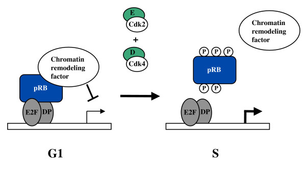
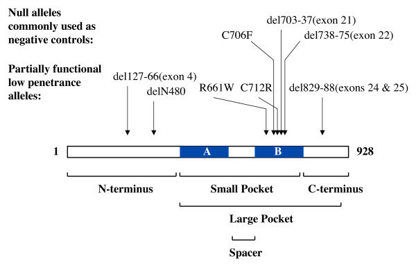
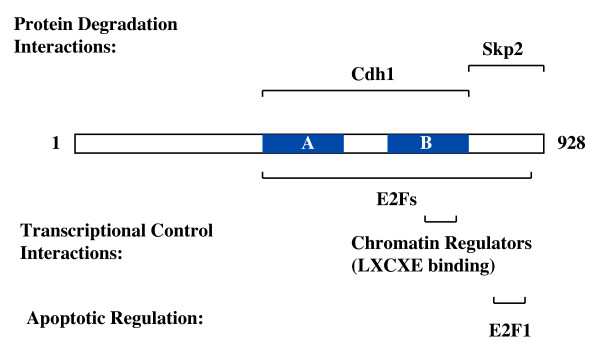
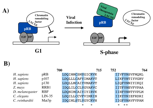
Biochemical analysis of the retinoblastoma protein's function has received considerable attention since it was cloned just over 20 years ago. During this time pRB has emerged as a key regulator of the cell division cycle and its ability to block proliferation is disrupted in the vast majority of human cancers. Much has been learned about the regulation of E2F transcription factors by pRB in the cell cycle. However, many questions remain unresolved and researchers continue to explore this multifunctional protein. In particular, understanding how its biochemical functions contribute to its role as a tumor suppressor remains to be determined. Since pRB has been shown to function as an adaptor molecule that links different proteins together, or to particular promoters, analyzing pRB by disrupting individual protein interactions holds tremendous promise in unraveling the intricacies of its function. Recently, crystal structures have reported how pRB interacts with some of its molecular partners. This information has created the possibility of rationally separating pRB functions by studying mutants that disrupt individual binding sites. This review will focus on literature that investigates pRB by isolating functions based on binding sites within the pocket domain. This article will also discuss the prospects for using this approach to further explore the unknown functions of pRB.


Figures

Figure 1
Figure 1
Transcriptional control of the cell cycle by pRB. In G1 pRB is bound to E2Fs and masks their ability to activate transcription. In turn, pRB can recruit a number of chromatin remodeling factors that can further inhibit the initiation of transcription. Mitogen signaling then activates cyclin/Cdk complexes that phosphorylate pRB in late G1 and early S-phase leading to the disassembly of this complex and the activation of transcription of genes needed for progression through S-phase.
Figure 2
Figure 2
Commonly used RB-1 mutations and pRB domain structure. The open reading frame of pRB is shown along with the locations of the A and B parts of the pocket. Arrows indicate the positions and alterations found in commonly studied low penetrance and high penetrance mutant alleles. Below the open reading frame, structural regions of pRB are identified along with their approximate locations.
Figure 3
Figure 3
Binding sites for E2Fs and other proliferative control activities. The open reading frame of pRB is shown with locations of the A and B parts of the pocket. Regions of contact that have been mapped for E2F transcription factors, chromatin regulators, and protein degradation machinery that are involved in controlling proliferation are diagramed.
Figure 4
Figure 4
pRB and the LXCXE motif. (A) A repressor complex composed of an E2F transcription factor, pRB, and a chromatin regulator is shown in G1. The interaction between pRB and the chromatin regulator is mediated by the peptide sequence LXCXE on the chromatin remodeling factor and the LXCXE binding cleft on pRB. Viral proteins like TAg, E7, and E1A can disrupt this complex because they contain an LXCXE motif. Disruption of this pRB-containing complex can advance the cell cycle into S-phase. (B) The LXCXE binding cleft on pRB is a shallow groove formed between two parallel helices. The amino acid sequence of these helices in human pRB is aligned to the analogous sequences from pRB-family proteins from humans, corn, fruit flies, worms, and algae. Shaded amino acids are identical between all protein comparisons. The asterisk denotes amino acids whose side chains make direct contact with LXCXE in the crystal structure reported by Lee et al. [38].
Figure 5
Figure 5
Post-translational modifications of pRB. The open reading frame of pRB is depicted with the A and B regions of the pocket indicated. Cyclin/Cdk phosphorylation sites that have been investigated by mutagenesis are shown as are sites of phosphorylation following DNA damage. The location of acetylated and sumoylated lysines are indicated. Arrows indicate the approximate location of Cdk4 and cyclin E binding sites that are needed for optimal phosphorylation of pRB. Caspase cleavage of pRB occurs on the C-terminal side of aspartate 886, removing amino acids 887 to 928.

Similar articles

Cited by

References

    1. Friend SH, Bernards R, Rogelj S, Weinberg RA, Rapaport JM, Albert DM, Dryja TP. A human DNA segment with properties of the gene that predisposes to retinoblastoma and osteosarcoma. Nature. 1986;323:643–646. doi: 10.1038/323643a0. - DOI - PubMed
    1. Sherr CJ. Cancer cell cycles. Science. 1996;274:1672–1677. doi: 10.1126/science.274.5293.1672. - DOI - PubMed
    1. Weinberg RA. The retinoblastoma protein and cell cycle control. Cell. 1995;81:323–330. doi: 10.1016/0092-8674(95)90385-2. - DOI - PubMed
    1. Dyson N. The regulation of E2F by pRB-family proteins. Genes Dev. 1998;12: 2245–2262. - PubMed
    1. Classon M, Harlow E. The retinoblastoma tumour suppressor in development and cancer. Nat Rev Cancer. 2002;2:910–917. doi: 10.1038/nrc950. - DOI - PubMed

LinkOut - more resources