

Ozone enhancement of lower airway allergic inflammation is prevented by gamma-tocopherol
- PMID: 17854713
- PMCID: PMC4465777
- DOI: 10.1016/j.freeradbiomed.2007.07.013
Ozone enhancement of lower airway allergic inflammation is prevented by gamma-tocopherol
Abstract
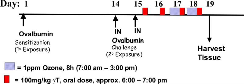
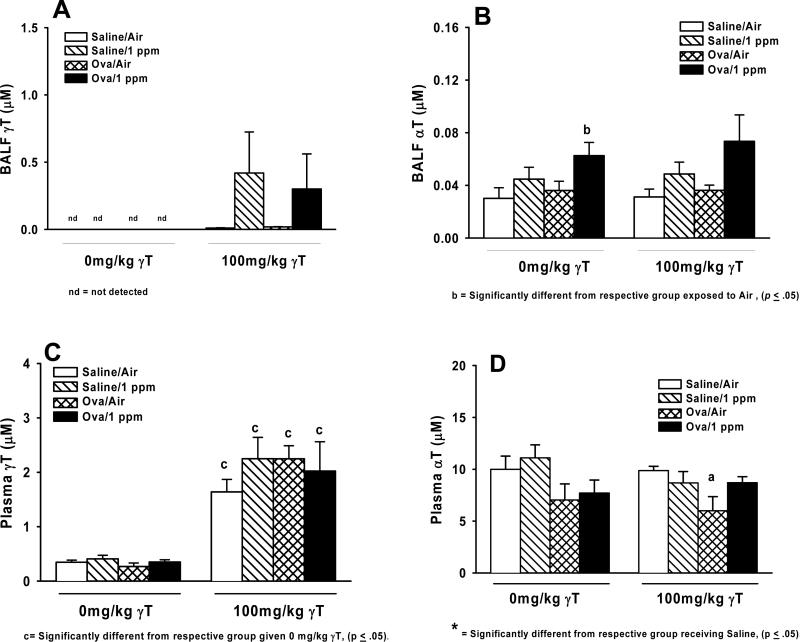
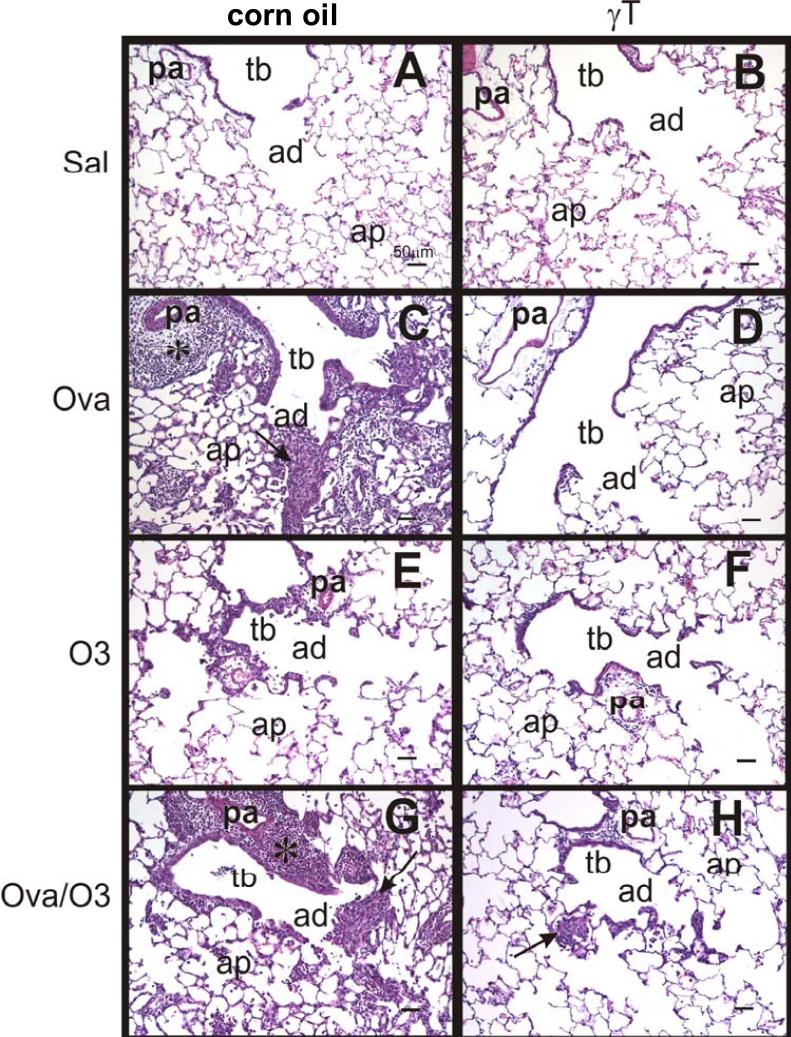
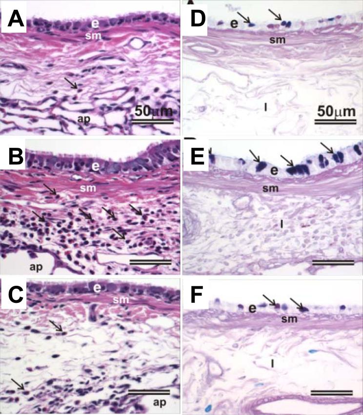
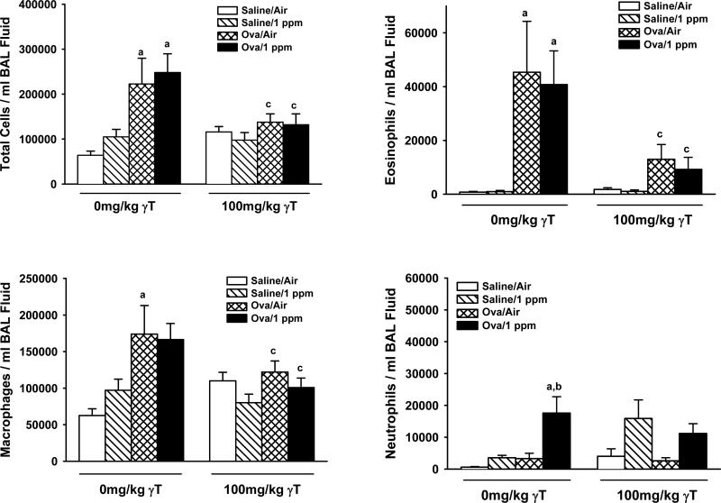
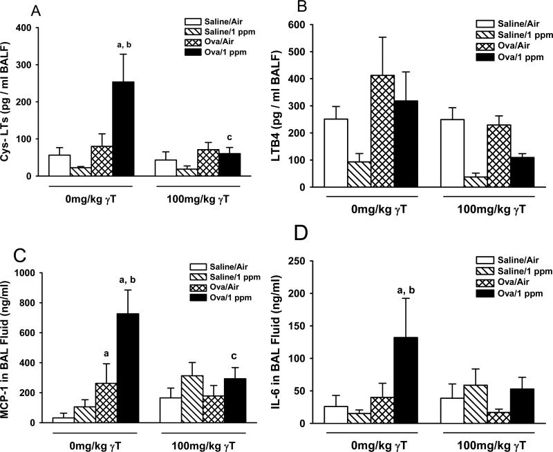
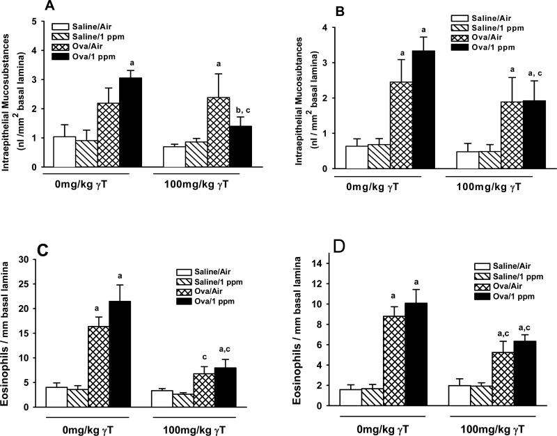
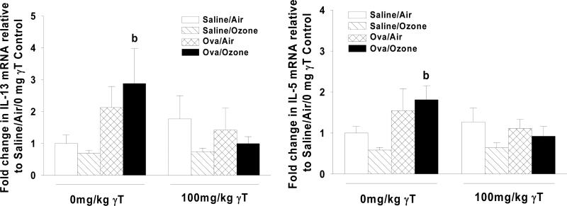
Ozone is a commonly encountered environmental oxidant which has been linked to asthma exacerbation in epidemiological studies. Ozone induces airway inflammation and enhances response to inhaled allergen. It has been suggested that antioxidant therapy may minimize the adverse effects of ozone in asthma. We have previously shown that the antioxidant gamma-tocopherol (gammaT), an isoform of vitamin E, also has anti-inflammatory effects. We employed a Brown Norway rat model of ozone-enhanced allergic responses to test the therapeutic effects of gammaT on O(3)-induced airway inflammation. Ovalbumin (OVA)-sensitized rats were intranasally challenged with 0 or 0.5% OVA on Days 1 and 2, and exposed to 0 or 1 ppm ozone (8 h/day) on Days 4 and 5. Rats were also given 0 or 100 mg/kg gammaT on Days 2 through 5. Pulmonary tissue and bronchoalveolar lavage fluid (BALF) were collected on Day 6. OVA challenge caused increased total cells (267% increase) and eosinophils (4000%) in BALF that was unaffected by ozone exposure. Morphometric evaluation of lung tissue revealed increases in intraepithelial mucosubstances (IM) (300%) and subepithelial eosinophils (400%) in main axial airways. Ozone exposure of allergic rats enhanced IM increases in proximal axial airways (200%), induced cys-leukotrienes, MCP-1, and IL-6 production in BALF, and upregulated expression of IL-5 and IL-13 mRNA. gammaT treatment had no effect on IM increases by allergen, but blocked enhancement by ozone. gammaT attenuated both OVA- or ozone-stimulated eosinophilic infiltration, and increases of BALF cys-leukotrienes, MCP-1, and IL-6, as well as IL-5 and IL-13 mRNA. These data demonstrate broad anti-inflammatory effects of a gammaT and suggest that it may be an effective therapy of allergic airway inflammation.
Figures
Similar articles
-
Gamma-tocopherol attenuates ozone-induced exacerbation of allergic rhinosinusitis in rats.Toxicol Pathol. 2009 Jun;37(4):481-91. doi: 10.1177/0192623309335630. Epub 2009 Apr 23. Toxicol Pathol. 2009. PMID: 19389874 Free PMC article.
-
Gamma-tocopherol prevents airway eosinophilia and mucous cell hyperplasia in experimentally induced allergic rhinitis and asthma.Clin Exp Allergy. 2008 Mar;38(3):501-11. doi: 10.1111/j.1365-2222.2007.02855.x. Epub 2007 Oct 26. Clin Exp Allergy. 2008. PMID: 17970781
-
Effects of concentrated ambient particles on normal and hypersecretory airways in rats.Res Rep Health Eff Inst. 2004 Aug;(120):1-68; discussion 69-79. Res Rep Health Eff Inst. 2004. PMID: 15543855
-
Effects of concentrated ambient particles and diesel engine exhaust on allergic airway disease in Brown Norway rats.Res Rep Health Eff Inst. 2009 Nov;(145):5-55. Res Rep Health Eff Inst. 2009. PMID: 20198910
-
Enhancement of nasal inflammatory and epithelial responses after ozone and allergen coexposure in Brown Norway rats.Toxicol Sci. 2002 Jun;67(2):284-94. doi: 10.1093/toxsci/67.2.284. Toxicol Sci. 2002. PMID: 12011488
Cited by
-
Short course gamma tocopherol did not mitigate effects of ozone on airway inflammation in asthmatics.Inhal Toxicol. 2020 Jun;32(7):279-281. doi: 10.1080/08958378.2020.1800147. Epub 2020 Jul 30. Inhal Toxicol. 2020. PMID: 32727225 Free PMC article. Clinical Trial.
-
Vitamin E isoforms as modulators of lung inflammation.Nutrients. 2013 Oct 31;5(11):4347-63. doi: 10.3390/nu5114347. Nutrients. 2013. PMID: 24184873 Free PMC article. Review.
-
Inhibition of myristoylated alanine-rich C kinase substrate (MARCKS) protein inhibits ozone-induced airway neutrophilia and inflammation.Exp Lung Res. 2010 Mar;36(2):75-84. doi: 10.3109/01902140903131200. Exp Lung Res. 2010. PMID: 20205598 Free PMC article.
-
Effects of ex vivo γ-tocopherol on airway macrophage function in healthy and mild allergic asthmatics.J Innate Immun. 2013;5(6):613-24. doi: 10.1159/000350234. Epub 2013 May 8. J Innate Immun. 2013. PMID: 23689260 Free PMC article.
-
Vascular cell adhesion molecule-1 expression and signaling during disease: regulation by reactive oxygen species and antioxidants.Antioxid Redox Signal. 2011 Sep 15;15(6):1607-38. doi: 10.1089/ars.2010.3522. Epub 2011 May 11. Antioxid Redox Signal. 2011. PMID: 21050132 Free PMC article. Review.
References
-
- Tolbert PE, Mulholland JA, MacIntosh DL, Xu F, Daniels D, Devine OJ, et al. Air quality and pediatric emergency room visits for asthma in Atlanta, Georgia, USA. Am J Epidemiol. 2000;151:798–810. - PubMed
-
- Gent JF, Triche EW, Holford TR, Belanger K, Bracken MB, Beckett WS, et al. Association of low-level ozone and fine particles with respiratory symptoms in children with asthma. Jama. 2003;290:1859–1867. - PubMed
-
- Kehrl HR, Peden DB, Ball B, Folinsbee LJ, Horstman D. Increased specific airway reactivity of persons with mild allergic asthma after 7.6 hours of exposure to 0.16 ppm ozone. J Allergy Clin Immunol. 1999;104:1198–1204. - PubMed
-
- Peden DB, Setzer RW, Jr., Devlin RB. Ozone exposure has both a priming effect on allergen-induced responses and an intrinsic inflammatory action in the nasal airways of perennially allergic asthmatics. Am J Respir Crit Care Med. 1995;151:1336–1345. - PubMed
Publication types
MeSH terms
Substances
Grants and funding
LinkOut - more resources
Full Text Sources
Miscellaneous
