Review
. 2008 Mar;1778(3):660-9.
doi: 10.1016/j.bbamem.2007.07.012. Epub 2007 Jul 27.

Adherens and tight junctions: structure, function and connections to the actin cytoskeleton

Affiliations
Review

Adherens and tight junctions: structure, function and connections to the actin cytoskeleton

Andrea Hartsock et al. Biochim Biophys Acta. 2008 Mar.

Abstract

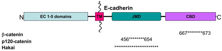
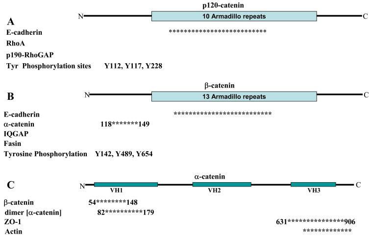
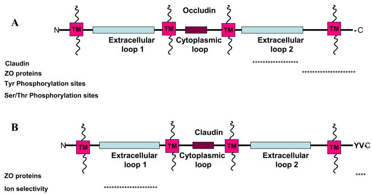
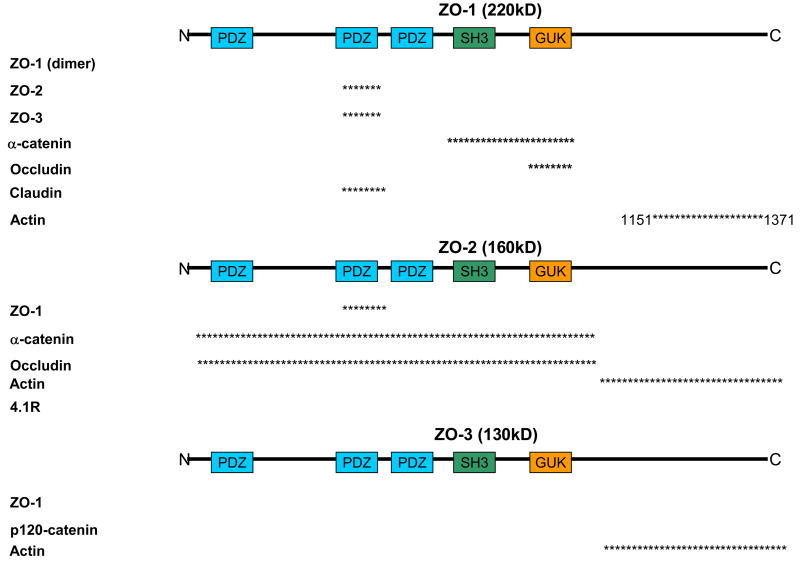
Adherens junctions and Tight junctions comprise two modes of cell-cell adhesion that provide different functions. Both junctional complexes are proposed to associate with the actin cytoskeleton, and formation and maturation of cell-cell contacts involves reorganization of the actin cytoskeleton. Adherens junctions initiate cell-cell contacts, and mediate the maturation and maintenance of the contact. Adherens junctions consist of the transmembrane protein E-cadherin, and intracellular components, p120-catenin, beta-catenin and alpha-catenin. Tight junctions regulate the paracellular pathway for the movement of ions and solutes in-between cells. Tight junctions consist of the transmembrane proteins occludin and claudin, and the cytoplasmic scaffolding proteins ZO-1, -2, and -3. This review discusses the binding interactions of the most studied proteins that occur within each of these two junctional complexes and possible modes of regulation of these interactions, and the different mechanisms that connect and regulate interactions with the actin cytoskeleton.


Figures

Figure 1
Figure 1
Adherens junctions are comprised of the single pass transmembrane protein, E-cadherin. The extracellular domain is proposed to form trans-interactions with E-cadherin on neighboring cells. The intracellular domain has two binding regions; juxtamembrane domain (JMD) and catenin binding domain (CBD). (TM; Transmembrane) The protein-protein interactions presented are limited to those involved in connections with the actin cytoskeleton. The asterisks represent the region proteins have been shown to bind. Not drawn to scale. Information was gathered based on mutational analysis or co-immunoprecipitation studies; all references are cited within the text.
Figure 2
Figure 2
Adherens junctions are comprised of A) p120-catenin, B) β-catenin, and C) α-catenin. All three proteins interact with additional proteins known to regulate actin cytoskeleton. Asterisks represent regions known to bind the respective protein. Numbers associated with the asterisks correspond to amino acids flanking the binding site. Not drawn to scale. Information was collected either through mutational analysis or co-immunoprecipitation. Proteins without asterisks represent interactions demonstrated by co-immunoprecipitation. References are cited within the text.
Figure 3
Figure 3
Tight Junction transmembrane proteins A) occludin and B) claudin, and proposed binding partners with corresponding binding regions. The asterisks represent the region in which the proteins have been shown to bind with the occludin and claudin. Not drawn to scale. Information was gathered based on mutational analysis or co-immunoprecipitation. All references are cited within the text.
Figure 4
Figure 4
ZO proteins are proposed to be a scaffolding protein that could link Tight junctions to the actin cytoskeleton through a direct interaction with actin or through additional protein interactions. In addition, ZO proteins may link Adherens junctions to Tight junctions through protein linkages. Proteins represented are proposed for these functions. Asterisks represent the region in which the proteins have been shown to bind with ZO protein. Numbers correspond with the amino acids on the parent ZO protein; not drawn to scale. Information was gathered based on mutational analysis or co-immunoprecipitation. Proteins without asterisks represent interactions demonstrated by co-immunoprecipitation but regions were not determined. All references are cited within the text.

References

    1. Gumbiner BM. Regulation of cadherin-mediated adhesion in morphogenesis. Nat Rev Mol Cell Biol. 2005;6:622–34. - PubMed
    1. Perez-Moreno M, Fuchs E. Catenins: keeping cells from getting their signals crossed. Dev Cell. 2006;11:601–12. - PMC - PubMed
    1. Capaldo CT, Macara IG. Depletion of E-cadherin disrupts establishment but not maintenance of cell junctions in Madin-Darby canine kidney epithelial cells. Mol Biol Cell. 2007;18:189–200. - PMC - PubMed
    1. Theard D, Steiner M, Kalicharan D, Hoekstra D, van Ijzendoorn SC. Cell polarity development and protein trafficking in hepatocytes lacking E-cadherin/beta-catenin-based adherens junctions. Mol Biol Cell. 2007;18:2313–21. - PMC - PubMed
    1. Schneeberger EE, Lynch RD. The tight junction: a multifunctional complex. Am J Physiol Cell Physiol. 2004;286:C1213–28. - PubMed

Publication types