Review
. 2008 Jan-Apr;96(1-3):421-51.
doi: 10.1016/j.pbiomolbio.2007.07.010. Epub 2007 Aug 10.

Late sodium current in failing heart: friend or foe?

Affiliations
Review

Late sodium current in failing heart: friend or foe?

Victor A Maltsev et al. Prog Biophys Mol Biol. 2008 Jan-Apr.

Abstract

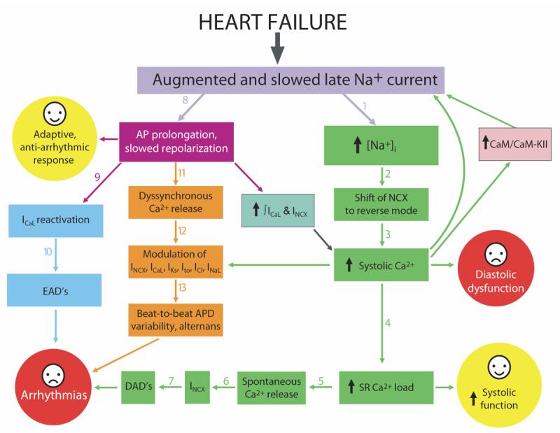
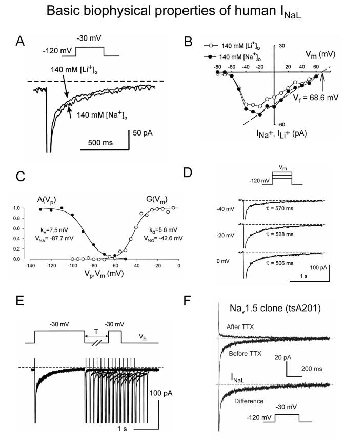
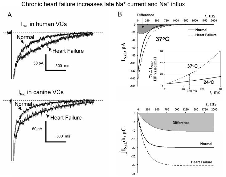
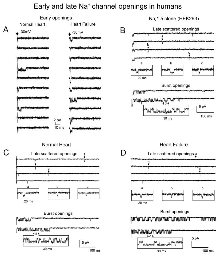
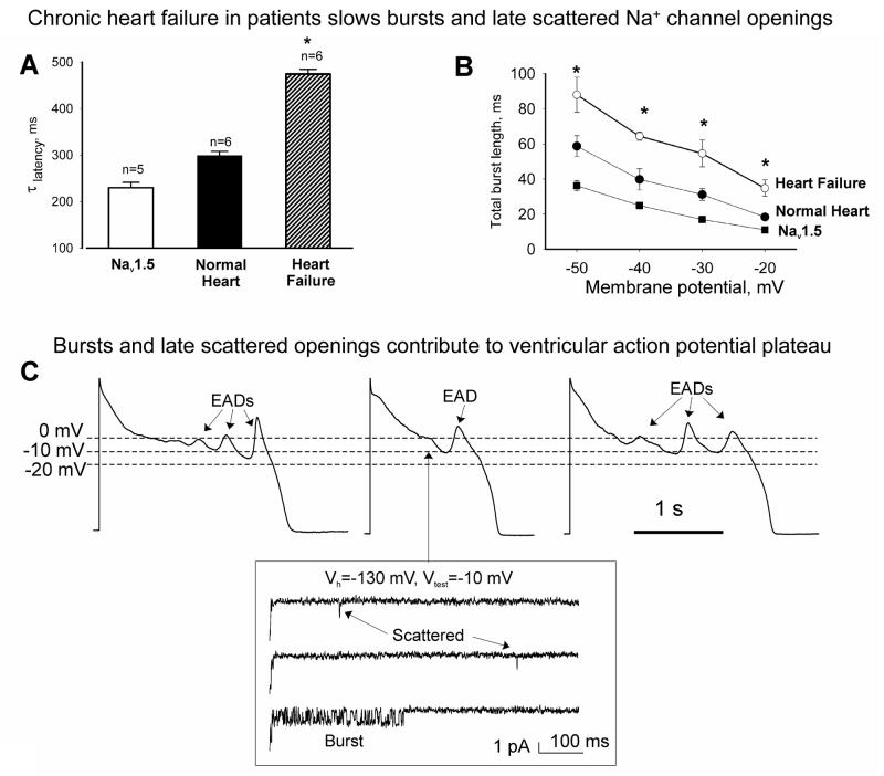
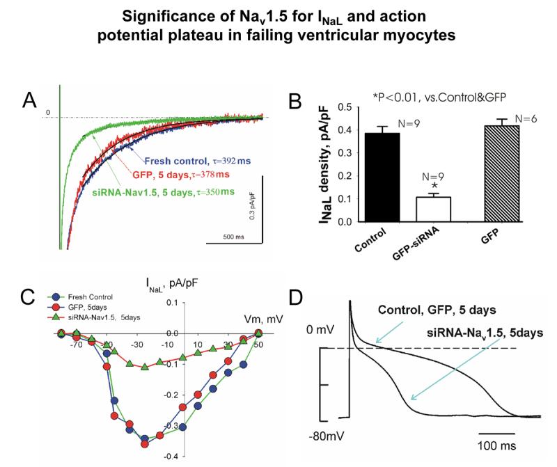
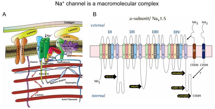
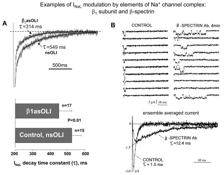
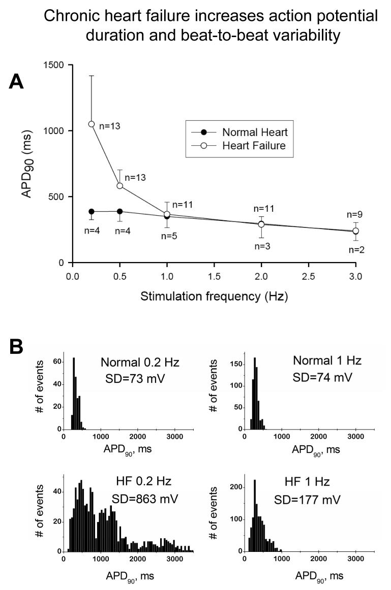
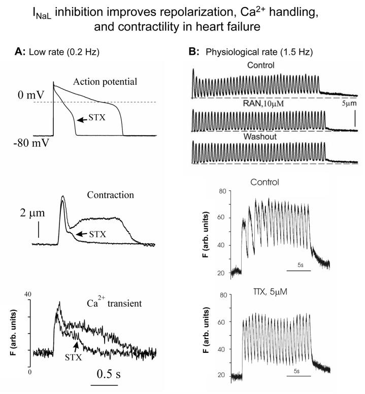
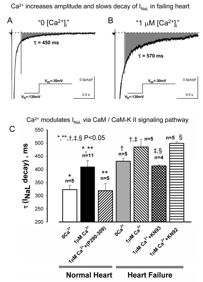
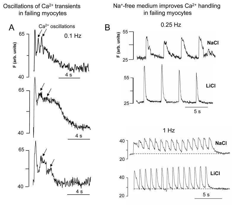
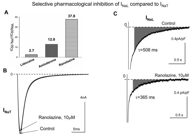
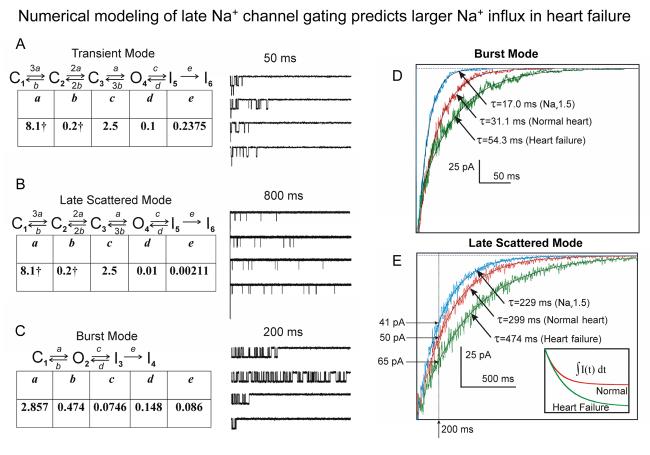
Most cardiac Na+ channels open transiently upon membrane depolarization and then are quickly inactivated. However, some channels remain active, carrying the so-called persistent or late Na+ current (INaL) throughout the action potential (AP) plateau. Experimental data and the results of numerical modeling accumulated over the past decade show the emerging importance of this late current component for the function of both normal and failing myocardium. INaL is produced by special gating modes of the cardiac-specific Na+ channel isoform. Heart failure (HF) slows channel gating and increases INaL, but HF-specific Na+ channel isoform underlying these changes has not been found. Na+ channels represent a multi-protein complex and its activity is determined not only by the pore-forming alpha subunit but also by its auxiliary beta subunits, cytoskeleton, calmodulin, regulatory kinases and phosphatases, and trafficking proteins. Disruption of the integrity of this protein complex may lead to alterations of INaL in pathological conditions. Increased INaL and the corresponding Na+ flux in failing myocardium contribute to abnormal repolarization and an increased cell Ca2+ load. Interventions designed to correct INaL rescue normal repolarization and improve Ca2+ handling and contractility of the failing cardiomyocytes. This review considers (1) quantitative integration of INaL into the established electrophysiological and Ca2+ regulatory mechanisms in normal and failing cardiomyocytes and (2) a new therapeutic strategy utilizing a selective inhibition of INaL to target both arrhythmias and impaired contractility in HF.


Figures

Figure 1
Figure 1
Integration of the late Na+ current into electrophysiological and Ca2+ regulatory mechanism in heart failure (see text for detail).
Figure 2
Figure 2
Biophysical properties of the slowly inactivating, late Na+ current (INaL) evaluated by whole cell patch clamp in human ventricular cardiomyocytes (A-E) and human cloned Nav1.5 expressed in tsA201 cells (F). A-B: Late current can be carried either by Na+ or Li+. B: I-V relation for INaL. C: examples of steady-state activation and availability curves, G(Vm) and A(Vp), respectively. D: Examples of original traces illustrating voltage-independent INaL decay. E: slow reactivation of INaL. F: INaL produced by Nav1.5 was assessed as difference current before application of a selective Na+ channel blocker tetrodotoxin (TTX, 30 μM) (20 averaged traces) and after TTX (14 averaged traces). Voltage protocols are shown at the traces. Recording was performed at 24°C. Reprinted from (Maltsev et al., 1998a) (A-E) and (Undrovinas et al., 2002)(F), used with permission.
Figure 3
Figure 3
A: Chronic heart failure slows and increases INaL. A: examples of whole cell INaL recordings in human and dog ventricular cardiomyocytes. B: Idealized INaL and their integrals in normal and failing dog cardiomyocytes of same size (200 pF) calculated using Q10 factors (37°C) and average parameters of INaL density and decay time constant. Larger and slower INaL in failing cardiomyocytes results in substantial increase in total charge (or Na+) transfer by INaL. Gray areas illustrate difference between failing and normal cells. Adapted from (Maltsev et al., 2007), used with permission.
Figure 4
Figure 4
Examples of Na+ channel activities that underlie peak (“early openings”, panel A) and late (panels B-D) Na+ currents in normal and failing human cardiomyocytes as well as in cardiac channel clone (Nav1.5) expressed in HEK293 cells. Note similarity between modes of late activity in normal hearts, failing hearts, and the clone. Cell-attached patches were held at −130 mV and step clamped to −30 mV at 23°C. Reprinted from (Undrovinas et al., 2002), used with permission from European Society of Cardiology.
Figure 5
Figure 5
Inactivation of both late scattered mode (A) or burst mode (B) of the late openings of Na+ channel was slowest in failing human cardiomyocytes compared with those from normal human hearts or heterologously expressed Nav1.5. *P<0.05, heart failure vs. normal heart or clone (Mean±SEM). Cell-attached patches: Vh = −140 mV, 24°C. (Reprinted from (Maltsev and Undrovinas, 2006) with permission from European Society of Cardiology). C: recordings of action potentials in failing human cardiomyocytes are shown along with late scattered mode and burst mode openings occurring at −10 mV, i.e. within the voltages of the action potential plateau. Reprinted from (Undrovinas et al., 2002), used with permission from European Society of Cardiology.
Figure 6
Figure 6
SCN5A gene regulates late sodium current (INaL) and action potential duration in failing heart. Silencing of SCN5A gene expression in ventricular cardiomyocytes of dogs with HF by adenovirally-transferred siRNA causes significant reduction in INaL density (A and B) in wide range of membrane potentials (C) and reduction of action potential duration (D). Shown are typical examples of INaL recordings, current-voltage relationships (whole cell patch clamp, 24°C) and action potential recordings (perforated patch, 35°C, 0.5 Hz pacing rate). Labels: “fresh control”- freshly isolated cells; “GFP” –cultured cells expressing reporter gene GFP, and “siRNA”- cultured cells with silenced SCN5A. Average data for action potential duration: fresh control, 515±51 ms, n=18; GFP control, 479±73 ms, n=8; siRNA, 181±17 ms, n=11; mean±SEM; n is number of cells; * P<0.01, vs. fresh control and GFP, ANOVA. From (Undrovinas et al., 2005).
Figure 7
Figure 7
Schematic illustration of Na+ channel as a macromolecular complex. A: The pore forming α subunit of the channel interacts with β-subunits, cytoskeleton and the extracellular matrix (Modified after (Nerbonne and Kass, 2005), used with permission). B: schematic presentation of the α subunit of the cardiac Na+ channel isoform (Nav1.5) with reported sites of interaction with β subunits (restricted only to β1 and β2) and other regulatory proteins.
Figure 8
Figure 8
Modulation of INaL by the channel environment elements: auxilliary β1-subunit and β-spectrin-based cytoskeleton. A: In normal dog ventricular myocytes, knocking down of SCN1B by antisense oligonucleotide (β1asOLI) significantly accelerates INaL decay compared to control nonsence oligonucleotide (nsOLI) (Undrovinas and Maltsev, 2002). B: Exposure of the cytoplasmic side of Na+ channel to the specific anti-β-spectrin antibody dramatically enhances activity of late Na+ channel openings culminating in the increased ensemble averaged late current. (rat ventricular myocytes, excised inside-out patch, from Undrovinas, Dubreuil and Makielski, unpublished)
Figure 9
Figure 9
A, Frequency-dependence of action potential duration in ventricular cardiomyocytes of normal dogs and dogs with chronic heart failure. Note that largest difference occurs at low pacing rates. B: at the low (0.2 Hz) and the physiologic (1 Hz) pacing rates, AP duration in failing myocytes exhibits significant beat-to beat variability (see respective SD values in the APD90 distribution histograms) Adapted from (Undrovinas et al., 1999) used with permission.
Figure 10
Figure 10
A: Examples of effects of a specific Na+ channel blocker saxitoxin (STX) on AP duration, contraction and Ca2+ transient in ventricular cardiomyocytes of dogs with chronic heart failure at a low pacing rate of 0.2 Hz STX reduces AP duration, abolishes “dome” phase of contraction and of Ca2+ transient in failing cells. B: At higher pacing rates a specific INaL blockers ranolazine reduces diastolic tension, and a specific Na+ channel blocker tetrodotoxin (TTX) reduces Ca2+ accumulation (Fluo-4 signals). Adapted from (Maltsev et al., 1998b; Undrovinas et al., 2006), used with permission.
Figure 11
Figure 11
Modulation of INaL by intracellular Ca2+ and CaM/CaM-Kinase signaling pathway in cardiomyocytes from normal and failing dog hearts. Elevated intracellular Ca2+ concentration up to 1 μM dramatically increases and slowes INaL. A, B: representative traces recorded in cardiomyocytes of failing hearts at “low” and “high” intracellular Ca2+. Gray areas indicate total integrals of INaL that proportionate to Na+ influx by INaL (beginning from 200 ms after depolarization onset depicted by the vertical bar). C: statistical data on INaL decay changes produced by perturbations of Ca2+/CaM/CaM-KII cascade in cardiomyocytes from normal and failing dog hearts. N corresponds to the number of cells tested. Adapted from (Maltsev et al., 2002a), used with permission.
Figure 12
Figure 12
A: Examples of fluctuations of Ca2+ transient (Fluo 4 signals) observed in ventricular myocytes of a canine chronic HF model at low pacing rates. B: Replacement of external Na+ by Li+ accelerates Ca2+ transients, abolishes Ca2+ oscillations, and decreases diastolic Ca2+ level in canine failing cardiomyocytes at low and high pacing rates, respectively (Undrovinas N., Sabbah H., Undrovinas A., unpublished). To avoid Li+ accumulation in the cells, the Na+-free solution was applied only for a few contraction cycles. Also, a new member NCLX of NCX superfamily has been recently discovered in a variety of tissues including heart (Palty et al., 2004). A distinct feature of NCLX is its ability to slowly transport Li+ in exchange to Ca2+, indicating that Li+ can be no longer considered as an “ideal” NCX blocker. Accordingly, if NCLX is functional in the failing canine myocytes, it could also prevent Li+ accumulation in the experimentation illustrated in panel B.
Figure 13
Figure 13
A: Comparison of the potencies of lidocaine, amiodarone and ranolazine to selectively inhibit INaL over the peak transient Na+ current (INaT). Shown are the ratios of IC50, i.e. the drug concentrations causing 50% resting block of the currents. The higher numbers correspond to the higher selectivity to block INaL. B, C: examples of a highly selective blockade of INaL over INaT produced by ranolazine in canine ventricular cardiomyocytes. Reprinted from (Undrovinas et al., 2006), used with permission.
Figure 14
Figure 14
A-C: Markov chain kinetic models describing the three major modes of Na+ channel gating in human cardiomyocytes: transient mode (A), late scattered openings (B) and bursts (C), together with simulated traces and transition rates; traces were simulated from 3, 5, and 1 channel(s), respectively. Total simulation times are indicated above the traces. D,E: The model predicts increased and slowed INaL in a failing human ventricular myocyte vs. a normal human myocyte and heterologously expressed Nav1.5 for BM (F) and LSM (G). Inset in panel E shows a substantially larger integral of the LSM current in a failing myocyte vs. a normal myocyte; the box size is 50 pC ×1990 ms. Shown are simulated cumulative activities of 20000 LSM channels or 193 BM channels. Channel numbers were chosen to correspond to a typical human ventricular myocyte. Reprinted from (Maltsev and Undrovinas, 2006) with permission from European Society of Cardiology.

References

    1. Abriel H, Kass RS. Regulation of the voltage-gated cardiac sodium channel Nav1.5 by interacting proteins. Trends Cardiovasc Med. 2005;15:35–40. - PubMed
    1. Ahern CA, Zhang JF, Wookalis MJ, Horn R. Modulation of the cardiac sodium channel NaV1.5 by Fyn, a Src family tyrosine kinase. Circ Res. 2005;96:991–998. Epub 2005 Apr 2014. - PubMed
    1. Akopian AN, Sivilotti L, Wood JN. A tetrodotoxin-resistant voltage-gated sodium channel expressedy sensory neurones. Nature. 1996;379:257–262. - PubMed
    1. Allouis M, Le Bouffant F, Wilders R, Peroz D, Schott JJ, Noireaud J, Le Marec H, Merot J, Escande D, Baro I. 14-3-3 is a regulator of the cardiac voltage-gated sodium channel Nav1.5. Circ Res. 2006;98:1538–1546. Epub 2006 May 1525. - PubMed
    1. Aman TK, Raman IM. Subunit dependence of Na channel open-channel block and slow inactivation in cerebellar neurons. Biophys J. 2006;22:22. - PMC - PubMed

Publication types