Review
. 2008;40(2):199-214.
doi: 10.1016/j.biocel.2007.07.015. Epub 2007 Aug 6.

Bridging structure with function: structural, regulatory, and developmental role of laminins

Affiliations
Review

Bridging structure with function: structural, regulatory, and developmental role of laminins

Julia Tzu et al. Int J Biochem Cell Biol. 2008.

Abstract

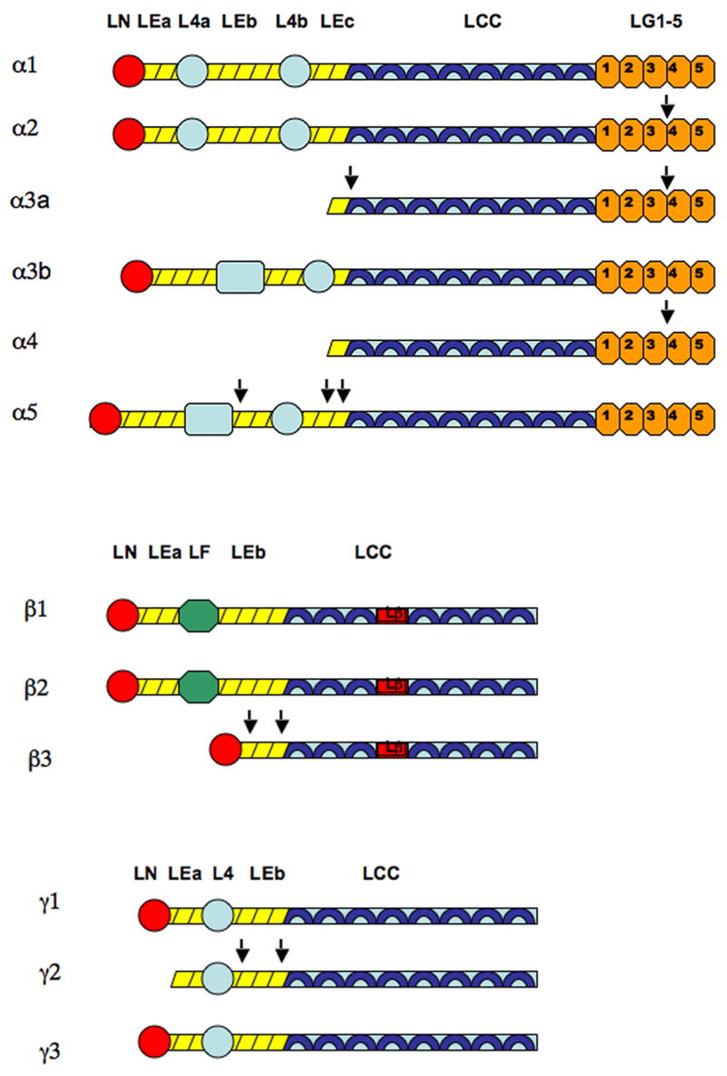
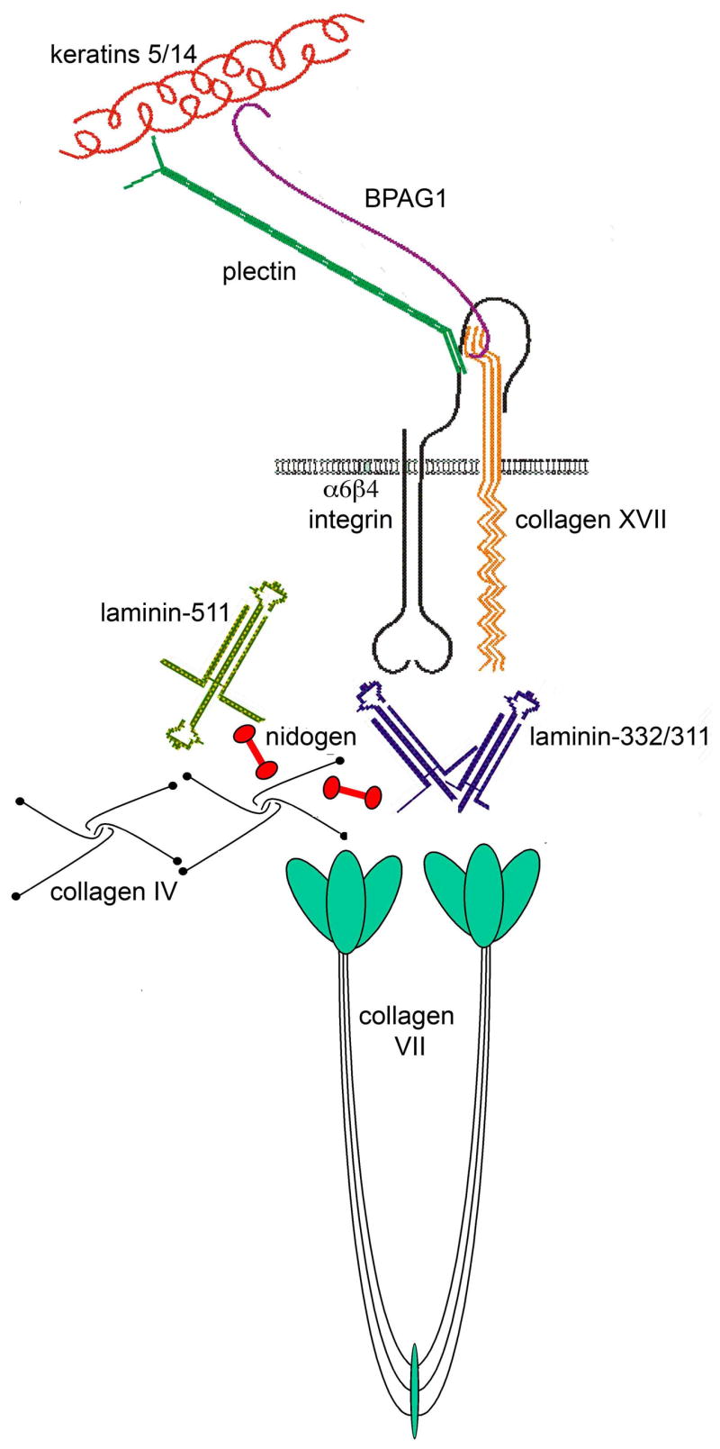
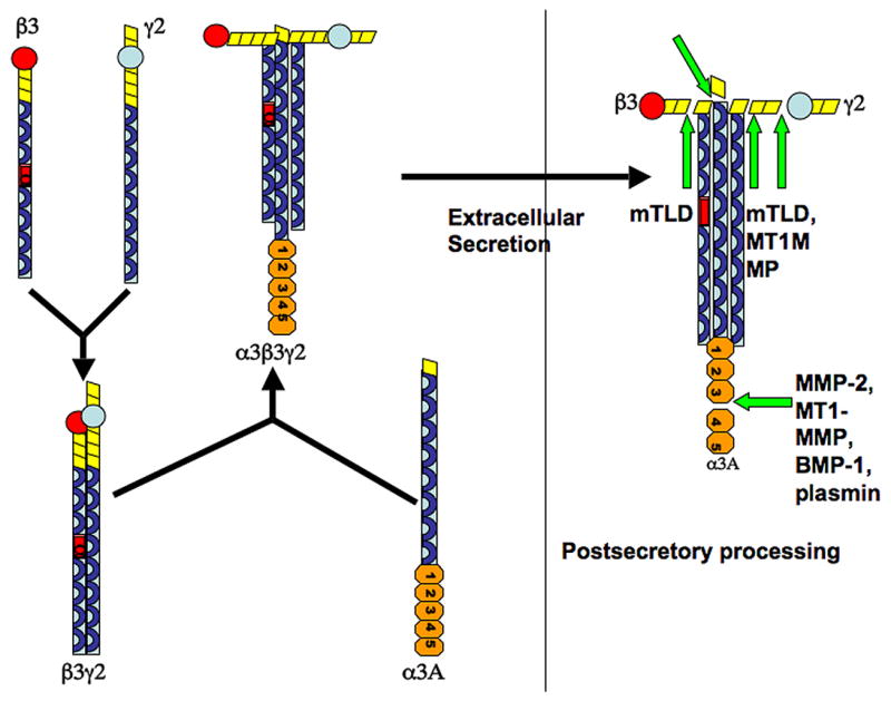
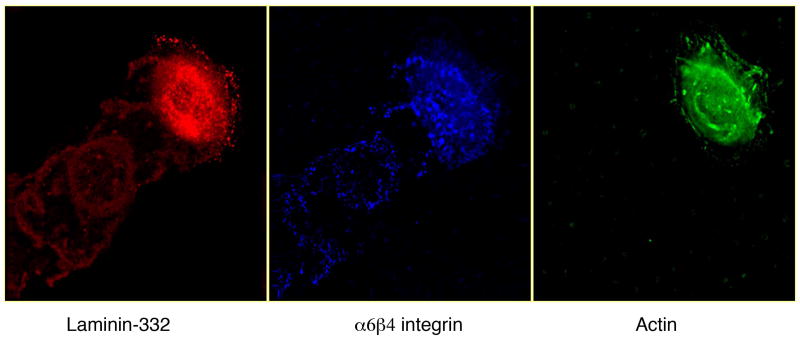
The basement membrane is a highly intricate and organized portion of the extracellular matrix that interfaces with a variety of cell types including epithelial, endothelial, muscle, nerve, and fat cells. The laminin family of glycoproteins is a major constituent of the basement membrane. The 16 known laminin isoforms are formed from combinations of alpha, beta, and gamma chains, with each chain containing specific domains capable of interacting with cellular receptors such as integrins and other extracellular ligands. In addition to its role in the assembly and architectural integrity of the basement membrane, laminins interact with cells to influence proliferation, differentiation, adhesion, and migration, processes activated in normal and pathologic states. In vitro these functions are regulated by the post-translational modifications of the individual laminin chains. In vivo laminin knockout mouse studies have been particularly instructive in defining the function of specific laminins in mammalian development and have also highlighted its role as a key component of the basement membrane. In this review, we will define how laminin structure complements function and explore its role in both normal and pathologic processes.


Figures

Figure 1
Figure 1
Domains of laminin alpha, beta, and gamma chains. Short arms of each chain are comprised of rodlike EGF-like tandems (LEa, LEb, LEc) and globular domains (LN, L4a, L4b, L4, LF). Long arms of each chain are comprised of alpha helical coils (LCC). Beta chains have a characteristic interruption of the coiled-coiled structure, known as the laminin β-knob domain (Lβ). Alpha chains also have a distinctive globular domain, consisting of five repeats (LG1-5), at the C terminal of the long arm (Patarroyo et al. 2002). Laminin 332 processing occurs in alpha, beta, and gamma chains. Note that processing occurs at both N and the LG3-G4 site of the C terminals in the alpha 3A chain (Aumailley et al. 2003). The α2 and α4 chains also undergo processing at the LG3-G4 site (Talts et al. 1998; Talts et al. 2000). The α5 chain undergoes N terminal processing at multiple sites within the EGF-like domains (Bair et al. 2005). The β3 chain undergoes processing at two sites along its short arm (Nakashima et al. 2005). Processing of the γ chain occurs at two sites as well, producing an internal DIII fragment that is implicated in cell motility (Koshikawa et al. 2005; Veitch et al. 2003). Cleavage sites are indicated by arrows.
Figure 2
Figure 2
Structure and composition of dermal-epidermal basement membrane zone. Laminin 511 and collagen IV constitute the major polymeric networks of the lamina densa, connected by nidogens as well as interactions with heparan sulfate proteoglycans such as perlecan. Laminin 332 is a component of the hemidesmosome-anchoring filament complex, bridging the cell surface with the dermis. It is linked to the rest of the ECM through its interactions with type VII collagen and laminin 311.
Figure 3
Figure 3
Assembly of laminin heterotrimers. Shown here is the assembly of the laminin 332 heterotrimer. Individual chains have all been glycosylated prior to dimmer/trimer assembly. Assembly begins first by the stable association of the β and γ chains. The α chain then combines with this dimer in order for the trimeric molecule to be secreted out of the cell. Addition of the α chain is the rate limiting step (Schneider et al. 2006). Proteolytic processing occurs after the trimer is secreted extracellularly.
Figure 4
Figure 4
Laminin 332 is a major component of the extracellular matrix of many epithelial cells. Shown here by triple label indirect immunofluorescence microscopy, laminin 332 localizes to trails behind migrating human keratinocytes along with its ligand α6β4 integrin. Actin staining is also included to indicate position of the cell.

References

    1. Amano S, Scott IC, Takahara K, Koch M, Champliaud MF, Gerecke DR, Keene DR, Hudson DL, Nishiyama T, Lee S, Greenspan DS, Burgeson RE. Bone morphogenetic protein 1 is an extracellular processing enzyme of the laminin 5 gamma 2 chain. J Biol Chem. 2000;275:22728–22735. - PubMed
    1. Ashton GH, Sorelli P, Mellerio JE, Keane FM, Eady RA, McGrath JA. Alpha 6 beta 4 integrin abnormalities in junctional epidermolysis bullosa with pyloric atresia. Br J Dermatol. 2001;144:408–414. - PubMed
    1. Aumailley M, Bruckner-Tuderman L, Carter WG, Deutzmann R, Edgar D, Ekblom P, Engel J, et al. A simplified laminin nomenclature. Matrix Biol. 2005;24:326–332. - PubMed
    1. Aumailley M, El Khal A, Knoss N, Tunggal L. Laminin 5 processing and its integration into the ECM. Matrix Biol. 2003;22:49–54. - PubMed
    1. Bader BL, Smyth N, Nedbal S, Miosge N, Baranowsky A, Mokkapati S, Murshed M, Nischt R. Compound genetic ablation of nidogen 1 and 2 causes basement membrane defects and perinatal lethality in mice. Mol Cell Biol. 2005;25:6846–6856. - PMC - PubMed

MeSH terms

LinkOut - more resources