. 2007 Nov;18(11):4591-602.
doi: 10.1091/mbc.e07-02-0157. Epub 2007 Sep 12.

Dual actin-bundling and protein kinase C-binding activities of fascin regulate carcinoma cell migration downstream of Rac and contribute to metastasis

Affiliations

Dual actin-bundling and protein kinase C-binding activities of fascin regulate carcinoma cell migration downstream of Rac and contribute to metastasis

Yosuke Hashimoto et al. Mol Biol Cell. 2007 Nov.

Abstract

Recurrence of carcinomas due to cells that migrate away from the primary tumor is a major problem in cancer treatment. Immunohistochemical analyses of human carcinomas have consistently correlated up-regulation of the actin-bundling protein fascin with a clinically aggressive phenotype and poor prognosis. To understand the functional and mechanistic contributions of fascin, we undertook inducible short hairpin RNA (shRNA) knockdown of fascin in human colon carcinoma cells derived from an aggressive primary tumor. Fascin-depletion led to decreased numbers of filopodia and altered morphology of cell protrusions, decreased Rac-dependent migration on laminin, decreased turnover of focal adhesions, and, in vivo, decreased xenograft tumor development and metastasis. cDNA rescue of fascin shRNA-knockdown cells with wild-type green fluorescent protein-fascin or fascins mutated at the protein kinase C (PKC) phosphorylation site revealed that both the actin-bundling and active PKC-binding activities of fascin are required for the organization of filopodial protrusions, Rac-dependent migration, and tumor metastasis. Thus, fascin contributes to carcinoma migration and metastasis through dual pathways that impact on multiple subcellular structures needed for cell migration.


Figures
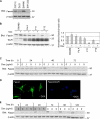
Figure 1.
Figure 1.
Development of inducible fascin-knockdown SW480 cells. (A) Immunoblot for fascin expression in human colon carcinoma cell lines. C2C12 skeletal myoblasts were used as a normal cell control. (B) Development of SW480 cells expressing TetR (PaA) and inducible fascin-knockdown clones F11 and F12. Doxycycline was applied at 1 μg/ml for 72 h. Graph shows normalized fascin levels (±SD) by scanning densitometry from triplicate experiments. (C) Time and concentration dependence of fascin knockdown in IKD-F11 cells. (D) Indirect immunofluorescent staining for fascin, in IKD-F11 cells maintained in the absence or presence of 0.5 μg/ml doxycycline for 72 h. Bar, 20 μm. Inset, detail of fascin-containing protrusions and associated filopodia. Bar, 10 μm. (E) Persistence of fascin knockdown. IKD-F11 cells were left untreated as controls or treated with the indicated concentrations of doxycycline for 48 h (shaded doxycycline panel), and then they were cultured in the absence of doxycycline for 72 h (unshaded, no doxycycline). The total experimental time was 120 h. Replicate cultures were lysed at the indicated timepoints and the levels of fascin protein examined by immunoblot during and after doxycycline treatment. Fascin knockdown in samples that had received doxycycline persisted for 72 h. Molecular mass markers are in kilodaltons.
Figure 2.
Figure 2.
Altered morphology of protrusions and loss of filopodia in fascin-knockdown cells. (A) Phase contrast views of SW480-Pa and IKD-F11 cells after 48 h culture in the absence or presence of 0.5 μg/ml doxycycline and plating for 2 h on 15 nM LN. Bar, 50 μm. (B) Scanning electron micrographs of IKD-F11 Fas+ cells adherent on LN. Examples of protrusions are arrowed (left), with enlarged view of the protrusive tips (right). Bars, 10 μm. (C) Scanning electron micrographs of IKD-F11 Fas− cells adherent on LN. Examples of residual protrusions are arrowed (left), with enlarged view of the tips (right). Bars, 10 μm. (D) F-actin organization in IKD-F11 cells adherent on LN under Fas+ and Fas− conditions. Bar, 20 μm. (E) IKD-F11 Fas− cells have decreased numbers of filopodia. Box and whisker plot of the number of filopodia per cell, quantified from at least 26 phalloidin-stained cells in three independent experiments.
Figure 3.
Figure 3.
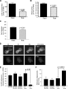
Fascin knockdown decreases cell migration and focal adhesion turnover. (A) transfilter migration of IKD-F11 Fas+ and Fas− cells. Each column is the mean of triplicate experiments; bars indicate SEM. (B) Haptokinetic migration of IKD cells on 15 nM LN. Migration speeds were calculated from time-lapse movies by measurements of the change in position of cell nuclei over time. Each column is the mean of three experiments; bars indicate SEM. See also Supplemental Movies 1 and 2. (C) Directional persistence of IKD cells migrating on LN. Net displacement of cell bodies was calculated from time-lapse movies in the Openlab program. Each column is the mean of three experiments; bars indicate SEM. (D) IKD-F11 cells under Fas+ and Fas− conditions and transiently expressing mRFP-paxillin were plated on 15 nM LN for 4 h. mRFP-paxillin–containing focal adhesions were imaged using a CARV spinning disk microscope by time-lapse imaging. Still images from each time-lapse series are shown at selected time points. Bars, 10 μm. See also Supplemental Movies 3 and 4. (E) Percentage of total adhesions disassembled over time by SW480 and F11 cells under the indicated conditions was calculated in ImageJ. Each column represents mean data from four cells per treatment and three independent experiments; bars indicate SEM. (F) The mean time taken for adhesions to disappear in SW480Pa and IKD-F11 cells, either in the absence or presence of doxycycline, was calculated from measuring 12–15 adhesions from five cells from three independent experiments. Each column represents the mean; bars indicate SEM.
Figure 4.
Figure 4.
Migration of fascin-knockdown cells is insensitive to Rac inhibition. (A) Migration speeds of IKD-F11 Fas+ cells (lanes 1–4) and IKD-F11 Fas− cells (lanes 5–8) adherent on 15 nM LN were calculated from time-lapse movies under control conditions (1, 4) or after pretreatment with 1 μM BIM (2, 6), 10 μM Y27632 (3, 7), or 100 μM NSC23766 (4, 8). Columns 1 and 4 represent the mean of nine independent experiments; other columns are means of three experiments. Bars indicate SEM, and p values are shown on the figure. See also Supplemental Movies 5 and 6. (B) Rac immunoblot to show that Rac protein levels were unchanged upon fascin depletion. Cell lysates were prepared from cells adhered to 15 nM LN for 2 h. (C) Rac activity is equivalent in LN-adherent IKD-F11 Fas+ and Fas− cells. Controls were constitutively active (CA) Rac1 protein and C2C12 cells in suspension or adhered to 50 nM FN for 15 min. Each column represents the mean of three experiments; bars indicate SEM.
Figure 5.
Figure 5.
Properties of IKD-F11 cells rescued with wild-type or mutant fascins. (A) F-actin organization in the cell lines under different experimental conditions as indicated. All cells were plated on 15 nM LN for 2 h. NSC23766 treatment was 100 μM for 18 h. Bars, 20 μm. Representative of three independent experiments. (B) quantification of cell morphologies in the absence or presence of rescue with wild-type or mutant fascins. Cell morphologies were scored in three categories: round [R], intermediate [I] and flat [F] (examples of the categories are labeled in A). Each column represents mean data from >100 cells derived from two to four independent experiments; bars indicate SEM. (C) Effect of Rac inhibition on directional persistence of GFP-XtfasS33D and GFP-XtfasS33A cells. The net displacement of cells migrating on 15 nM LN for 200 min, without or with treatment with 100 μm NSC23766, was calculated from time-lapse movies. Each column represents the mean from at least 30 cells, bars indicate SEM. See also Supplemental Movies 7 and 8.
Figure 6.
Figure 6.
Fascin depletion decreases xenograft tumor development and metastasis in nude mice. (A) Immunoblot for fascin demonstrates that fascin protein levels in the subcutaneous tumors of nude mice given doxycycline in their drinking water were specifically reduced in the mice inoculated with IKD-F11 cells (F11+ Dox). Each lane represents the extract of a single tumor harvested after 32 d; three or four samples are shown for each experimental condition. Extracts of C2C12 cells (C2) were included as positive controls. (B) Immunohistochemical staining for human fascin demonstrates the specific and uniform depletion of fascin protein within the IKD-F11 tumors of nude mice given doxycycline. (C) Metastatic potential of IKD-F11 cells is reduced in nude mice given doxycycline (Fas− condition). Each column represents the mean of at least 6 mice; bars indicate SEM.

Similar articles

Cited by

References

    1. Adams J. C. Characterization of cell-matrix adhesion requirements for the formation of fascin microspikes. Mol. Biol. Cell. 1997;8:2345–2363. - PMC - PubMed
    1. Adams J. C. Roles of fascin in cell adhesion and motility. Curr. Opin. Cell Biol. 2004;16:590–596. - PubMed
    1. Adams J. C., Clelland J. D., Collett G. D., Matsumura F., Yamashiro S., Zhang L. Cell-matrix adhesions differentially regulate fascin phosphorylation. Mol. Biol. Cell. 1999;10:4177–4190. - PMC - PubMed
    1. Adams J. C., Schwartz M. A. Stimulation of fascin spikes by thrombospondin-1 is mediated by the GTPases Rac and Cdc42. J. Cell Biol. 2000;150:807–822. - PMC - PubMed
    1. Akiyama S. K., Yamada S. S., Chen W. T., Yamada K. M. Analysis of fibronectin receptor function with monoclonal antibodies: roles in cell adhesion, migration, matrix assembly, and cytoskeletal organization. J. Cell Biol. 1989;109:863–875. - PMC - PubMed

Publication types

MeSH terms

LinkOut - more resources