. 2007 Nov;81(22):12323-36.
doi: 10.1128/JVI.01506-07. Epub 2007 Sep 12.

Localization and membrane topology of coronavirus nonstructural protein 4: involvement of the early secretory pathway in replication

Affiliations

Localization and membrane topology of coronavirus nonstructural protein 4: involvement of the early secretory pathway in replication

M Oostra et al. J Virol. 2007 Nov.

Abstract

The coronavirus nonstructural proteins (nsp's) derived from the replicase polyproteins collectively constitute the viral replication complexes, which are anchored to double-membrane vesicles. Little is known about the biogenesis of these complexes, the membrane anchoring of which is probably mediated by nsp3, nsp4, and nsp6, as they contain several putative transmembrane domains. As a first step to getting more insight into the formation of the coronavirus replication complex, the membrane topology, processing, and subcellular localization of nsp4 of the mouse hepatitis virus (MHV) and severe acute respiratory syndrome-associated coronavirus (SARS-CoV) were elucidated in this study. Both nsp4 proteins became N glycosylated, while their amino and carboxy termini were localized to the cytoplasm. These observations imply nsp4 to assemble in the membrane as a tetraspanning transmembrane protein with a Nendo/Cendo topology. The amino terminus of SARS-CoV nsp4, but not that of MHV nsp4, was shown to be (partially) processed by signal peptidase. nsp4 localized to the endoplasmic reticulum (ER) when expressed alone but was recruited to the replication complexes in infected cells. nsp4 present in these complexes did not colocalize with markers of the ER or Golgi apparatus, while the susceptibility of its sugars to endoglycosidase H indicated that the protein had also not traveled trough the latter compartment. The important role of the early secretory pathway in formation of the replication complexes was also demonstrated by the inhibition of coronaviral replication when the ER export machinery was blocked by use of the kinase inhibitor H89 or by expression of a mutant, Sar1[H79G].


Figures

FIG. 1.
FIG. 1.
Schematic representation of the coronavirus pp1ab polyprotein and of the nsp4 constructs used in this study. The coronavirus pp1ab precursor is shown at the top. The mature cleavage products (nsp's) are indicated by numbers. The transition between ORF1a and ORF1b is indicated as a ribosomal frame shift (RFS), while arrowheads represent sites that are cleaved by the nsp3-encoded PLpro protein (gray), of which there are two in MHV and only one in SARS-CoV, or by the nsp5-encoded Mpro protein (black). Within the nsp's, key replicase domains have been highlighted. These include putative transmembrane domains (TM) and the ORF1b-encoded domains: RNA-dependent RNA polymerase (RdRp), Helicase (Hel), exonuclease (ExoN), endoribonuclease (N), and methyltransferase (MT). The different nsp4 fusion proteins used in this study are schematically depicted below. nsp4 is shown in gray, with the hydrophobic domains in white, while asterisks indicate the approximate locations of the potential N glycosylation sites (NXS/T) in MHV nsp4 and the triangle indicates the approximate location of the atypical glycosylation motif (NXC) in SARS-CoV nsp4. The N (MN)- and C (EGFP and HA)-terminal tags are also indicated.
FIG. 2.
FIG. 2.
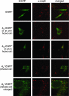
Localization of transiently expressed nsp4. LR7 cells, transfected with EGFP-, MHV nsp4-EGFP (4m-EGFP)-, or MHV nsp4′-EGFP (4m′-EGFP)-encoding constructs, were infected with MHV-A59. Cells were fixed at 6 h postinfection and processed for immunofluorescence microscopy using anti-nsp8 (α-nsp8) serum and a Cy3-conjugated antiserum to detect the MHV replication sites, as described in Materials and Methods. The second row shows a cell that is transfected (EGFP positive) but not infected (nsp8 negative), between cells that are infected (nsp8 positive) but not transfected (EGFP negative), whereas the lower rows show cells that are both transfected and infected (EGFP and nsp8 positive). At the right, a merged image of the α-nsp8 and the EGFP signal is shown. The bottom pictures are enlargements of the pictures in the row just above them.
FIG. 3.
FIG. 3.
Processing of MHV and SARS-CoV nsp4. vTF7-3-infected OST7-1 cells were transfected with the indicated constructs. The cells were labeled with 35S-labeled amino acids from 5 to 6 h postinfection, lysed, and processed for immunoprecipitation with specific antibodies, followed by SDS-10% PAGE. (A) Cells were transfected with MHV or SARS-CoV nsp4-EGFP-encoding constructs (4m-EGFP or 4s-EGFP, respectively) in the presence (+) or absence (−) of tunicamycin (TM). The same constructs were also in vitro transcribed and translated using the TNT coupled reticulocyte lysate system from Promega (ivt). Immunoprecipitations were performed with rabbit antiserum against the EGFP tag. (B) Cells were transfected with constructs encoding SARS-CoV nsp4-HA (4s-HA), a SARS-CoV nsp4-HA fusion protein containing a mutation of the NIC glycosylation motif (4s−glyc-HA) or an N-terminally tagged SARS-CoV nsp4-HA fusion protein (4s′-HA). The construct encoding SARS-CoV nsp4-HA (4s-HA) was also in vitro transcribed and translated using the TNT coupled reticulocyte lysate system from Promega (ivt). Immunoprecipitations were performed with rabbit serum against the HA tag, after which the samples were mock (−) or PNGaseF (+) treated. (C) The N-terminally tagged SARS-CoV nsp4-HA fusion protein was in vitro translated using the TNT coupled reticulocyte lysate system from Promega in the absence (−) or presence (+) of microsomal membranes (mm). (D) Cells were transfected with SARS-CoV nsp4 containing an N-terminal MN tag and a C-terminal HA tag (4s′-HA). Immunoprecipitation with rabbit antiserum against the HA tag was followed, after boiling of the sample, by a second immunoprecipitation with either J1.3 antiserum against the N-terminal tag (J1.3; left lane) or the antiserum against the C-terminal HA tag (α-HA; right lane). The positions and masses (in kDa) of the molecular-mass protein markers are indicated, while the numbers 1, 2, and 3 indicate different SARS-CoV nsp4 species, of which the number 1 species are modified by N-linked sugars. The two arrows in panel D point to species 1′ and 2′, which are protein species containing the amino-terminal tag. Only the relevant portions of the gels are shown.
FIG. 4.
FIG. 4.
O glycosylation of N-terminally tagged MHV and SARS-CoV nsp4. vTF7-3-infected OST7-1 cells were transfected with constructs encoding MHV nsp4-EGFP with or without the N-terminal tag (4m′-EGFP or 4m-EGFP, respectively), N-terminally tagged SARS-CoV nsp4-HA (4s′-HA), or N-terminally tagged EAV M (EAV M+9A) in the presence (+) or absence (−) of tunicamycin (TM) and/or brefeldin A (BFA). The cells were labeled with 35S-labeled amino acids from 5 to 6 h postinfection, and cell lysates were processed for immunoprecipitation with rabbit antiserum against EGFP or HA or, for EAV M, with J1.3, followed by SDS-10% PAGE. The positions of the molecular-mass protein markers are indicated on the left or right side of each gel. Only the relevant portions of the gels are shown.
FIG. 5.
FIG. 5.
Topology of MHV and SARS-CoV nsp4. vTF7-3-infected OST7-1 cells were transfected with constructs encoding the proteins indicated at the left. The cells were fixed at 6 h postinfection and permeabilized with Triton X-100 (left 2 columns) or digitonin (right 2 columns). Immunofluorescence analysis was performed with the antibodies indicated above the pictures, while EGFP indicates the EGFP fluorescence itself. α-Mc, anti-Mc; α-EGFP, anti-EGFP; α-HA, anti-HA.
FIG. 6.
FIG. 6.
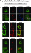
Recombinant MHV containing nsp4-EGFP. (A) A recombinant MHV containing the MHV nsp4-EGFP fusion gene at the position of the HE gene was generated. The wild-type (wt) and recombinant (rec) MHV genomes are schematically represented. Genes are indicated by numbers or letters. AAA indicates the poly(A) tail at the 3′end. (B, C) LR7 cells were infected with the recombinant virus, fixed at 6 h (B) or 8 h (C) postinfection, and stained with antibodies against marker proteins: anti-nsp8 (α-nsp8), anti-dsRNA (α-dsRNA), anti-calreticulin (α-calreticulin) (ER), or anti-GM130 (α-GM130) (Golgi apparatus). Merged images of the EGFP signal with the staining of the different markers are shown at the right. In each set, the lower pictures are enlargements of the images above. The white arrows in the upper part of panel C indicate nsp4-EGFP located at the replication sites.
FIG. 7.
FIG. 7.
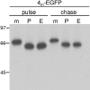
Maturation of the glycosylation of nsp4. LR7 cells were infected with the recombinant MHV containing the nsp4-EGFP fusion gene (4m-EGFP). The cells were labeled from 5 to 6 h postinfection (pulse), followed by a 90-min chase (chase), after which cell lysates were prepared and subjected to immunoprecipitation using the anti-EGFP antiserum. The immunoprecipitated material was treated with either PNGaseF (P) or EndoH (E) or was mock (m) treated. The numbers at the left indicate the positions of the molecular-mass protein markers. Only the relevant portion of the gel is shown.
FIG. 8.
FIG. 8.
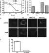
Role of the early secretory pathway in virus replication. LR-7 cells were infected with MHV-EFLM in the presence of H89, and the viral replication was determined by measuring luciferase expression. (A) The cells were incubated with different concentrations of H89 from 1 to 6 h postinfection, after which luciferase activity was determined (left y axis). The cell viability was determined by a WST-1 assay (right y axis). (B) MHV-EFLM-infected cells were lysed at 4 (first two bars) or 7 (last two bars) h postinfection, either with (second and fourth bars) or without (first and third bars) 3 h of incubation with 50 μM H89. (C) At 4 h postinfection, LR-7 cells were infected with MHV and fixed (most left panels) or incubation at 37°C was continued for 3 h in the absence (7 h) or presence of H89 (H89) or cycloheximide (CHX). After fixation, the cells were processed for immunofluorescence analysis using antibodies directed against dsRNA or nsp8. (D) LR-7 cells were transfected with plasmids expressing fusion proteins of YFP with either wild-type Sar1 or Sar1[H79G] and 24 h later infected with a recombinant MHV expressing RFP. At 16 h postinfection, the number of RFP-positive cells in the YFP-positive population was determined by flow cytometric analysis.
FIG. 9.
FIG. 9.
Schematic representation of the proposed MHV and SARS-CoV nsp4 topology and intracellular transport. (A) The topology of MHV and SARS-CoV nsp4 is presented, with both the N and C termini at the cytoplasmic side of the membrane. The four predicted transmembrane domains are shown as white boxes, the N-glycosylation sites are presented as asterisks, and the (partial) signal sequence cleavage of the SARS-CoV nsp4 is indicated by the dotted arrow. (B) Intracellular localization and cellular transport are shown for nsp4 and for the coronavirus M protein. The M protein is transported from the ER to the Golgi compartment via the intermediate compartment (IC), whereas nsp4 is transported to the DMVs without passing through the medial or trans cisternae of the Golgi compartment. The inhibitory effect of H89 treatment or expression of Sar1[H79G] on coronavirus replication is indicated.

Similar articles

Cited by

References

    1. Altan-Bonnet, N., R. Sougrat, and J. Lippincott-Schwartz. 2004. Molecular basis for Golgi maintenance and biogenesis. Curr. Opin. Cell Biol. 16:364-372. - PubMed
    1. Anand, K., J. Ziebuhr, P. Wadhwani, J. R. Mesters, and R. Hilgenfeld. 2003. Coronavirus main proteinase (3CLpro) structure: basis for design of anti-SARS drugs. Science 300:1763-1767. - PubMed
    1. Aridor, M., and W. E. Balch. 2000. Kinase signaling initiates coat complex II (COPII) recruitment and export from the mammalian endoplasmic reticulum. J. Biol. Chem. 275:35673-35676. - PubMed
    1. Baker, S. C., K. Yokomori, S. Dong, R. Carlisle, A. E. Gorbalenya, E. V. Koonin, and M. M. Lai. 1993. Identification of the catalytic sites of a papain-like cysteine proteinase of murine coronavirus. J. Virol. 67:6056-6063. - PMC - PubMed
    1. Belov, G. A., and E. Ehrenfeld. 2007. Involvement of cellular membrane traffic proteins in poliovirus replication. Cell Cycle 6:36-38. - PubMed

Publication types

MeSH terms

Substances