. 2007 Nov;81(22):12307-15.
doi: 10.1128/JVI.01002-07. Epub 2007 Sep 12.

Infection of cardiomyocytes and induction of left ventricle dysfunction by neurovirulent polytropic murine retrovirus

Affiliations

Infection of cardiomyocytes and induction of left ventricle dysfunction by neurovirulent polytropic murine retrovirus

Mohammed Khaleduzzaman et al. J Virol. 2007 Nov.

Abstract

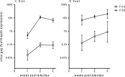
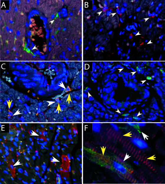
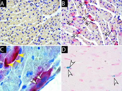
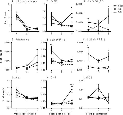
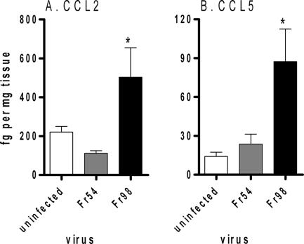
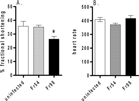
Viral infections of the heart are a causative factor of myocarditis as well as of sudden, unexpected deaths of children, yet the mechanisms of pathogenesis remain unclear, in part due to the relatively few animal models of virus-induced myocarditis. In the current study, we examined the ability of polytropic murine retroviruses to infect the heart and induce cardiac dysfunction. In situ hybridization and immunohistochemistry analysis detected virus-infected cardiomyocytes and macrophages in the heart. A significant decrease in left ventricle function, as measured by fractional shortening, was detected in mice infected with the neurovirulent retrovirus Fr98 but not in mice infected with the nonneurovirulent retrovirus Fr54. Virus infection was not associated with consistent findings of fibrosis or substantial cellular infiltrate. Fr98-induced left ventricle dysfunction was associated with a higher virus load, increased mRNA expression of the macrophage marker F4/80, increased chemokine production, and a small number of apoptotic cells in the heart.


Figures

FIG. 1.
FIG. 1.
Virus expression in (A) brain and (B) heart over 3 wpi in 129SvEv mice. Newborn mice were infected with 104 focus-forming units of Fr98 or Fr54 by intraperitoneal injection. Brain and heart tissue were removed from Fr98- and Fr54-infected mice at the indicated time. Virus gag RNA levels were determined using quantitative real-time PCR using SYBR green detection. The PCR product was sequenced to confirm the amplification of the correct gene. Data are expressed as virus mRNA levels as a percentage of Gapdh mRNA levels in each sample. Data are the means ± standard errors for four to eight samples per group per time point. Error bars indicate standard deviations.
FIG. 2.
FIG. 2.
Immunohistochemistry analysis of cell types in heart tissue. (A) Virus envelope protein (green fluorescence, white arrows) was detected on small cells located near blood vessels in Fr98-infected mice. (B) Macrophages were detected by Iba1 staining (red fluorescence, white arrows) near blood vessels in the heart. (C) Dual staining for virus envelope protein (red, white arrows) and macrophage marker Iba1 (green fluorescence, yellow arrows) show virus-infected macrophages (yellow fluorescence) in the heart. (D) XPR1-expressing cells in heart tissue. (E) Detection of virus envelope protein (red fluorescence) in multiple cells of the heart, including myocytes (white arrows). (F) Colocalization of virus envelope (green fluorescence, white arrows) and desmin (red fluorescence, yellow arrows) demonstrate virus infection of myocytes. Heart tissue from uninfected or Fr98-infected mice was removed at 18 to 21 dpi and processed for immunohistochemistry as described in Materials and Methods. Images were captured using Image Pro Plus 5.0 software. Magnification, ×200 (A, B, C, and D), ×100 (E), and ×1,000 (F).
FIG. 3.
FIG. 3.
(A to C) Confirmation of virus infection of myocytes by detection of viral RNA (red stain, white arrows) in myocytes. DIG-labeled RNA probe specific for the virus envelope gene was used to detect Fr98 viral RNA in (A) uninfected or (B and C) Fr98-infected mice. (D) Detection of TUNEL-positive apoptotic cells (blue nuclei, white arrows) near left ventricles of Fr98-infected mice. No TUNEL-positive cells were detected in heart tissue from uninfected or Fr54-infected mice. Heart tissue from uninfected or Fr98-infected mice was removed at 18 to 21 dpi and processed for in situ hybridization as described in Materials and Methods. Sections were counterstained with (A to C) hematoxylin or (D) neutral red. Images were captured using Image Pro Plus 5.0 software. Magnification, ×100 (A and B), ×1,000 (C), and ×100 (D).
FIG. 4.
FIG. 4.
mRNA expression of (A) α type 1 collagen, (B) F4/80, (C) beta 1 interferon, (D) gamma interferon, (E) Ccl4, (F) Ccl5, (G) Ccr1, (H) Ccr5, and (I) iNos in heart tissue of Fr98-infected, Fr54-infected, or uninfected 129SvEv mice. Newborn mice were infected with 104 focus-forming units of Fr98 or Fr54 by intraperitoneal injection. Heart tissue was removed from Fr98- and Fr54-infected mice at the indicated time, divided into two cross-sectional pieces, and snap-frozen in liquid nitrogen. mRNA levels were determined by using quantitative real-time PCR using SYBR green detection. Data were calculated as a percentage of Gapdh mRNA for each sample. Data are shown as the means ± standard errors (error bars) for four to seven samples per group per time point. Statistics were calculated using a two-way analysis of variance with Bonferroni's posttest. *, P < 0.05.
FIG. 5.
FIG. 5.
Protein expression of CCL2 and CCL5 in heart tissue from uninfected, Fr54-infected, and Fr98-infected mice. Mice were infected as described for Fig. 3. Heart tissue was homogenized in lysis buffer and analyzed for protein expression by mutliplex bead array. Data were calculated as femtograms of chemokine per milligram of tissue homogenate. Data are shown as the means ± standard errors (error bars) of five mice per group. Statistical analysis was completed using a one-way analysis of variance with a Newman-Keul's posttest. *, P < 0.05.
FIG. 6.
FIG. 6.
Echocardiogram analysis of left ventricular dysfunction in Fr98-infected mice. (A) Mean percentage of fractional shortening from Fr98-infected, Fr54-infected, or uninfected mice at 3 wpi (18 to 25 days postinfection). Data are the means ± standard errors (error bars) of six to nine mice per group and are the combined results from three separate experiments. (B) Mean heart rates of Fr98-infected, Fr54-infected, and uninfected mice at 3 wpi (18 to 25 days). Data are the means ± standard errors (error bars) of three to seven mice per group. *, P < 0.05.

Similar articles

Cited by

References

    1. Aukrust, P., T. Ueland, F. Muller, A. K. Andreassen, I. Nordoy, H. Aas, J. Kjekshus, S. Simonsen, S. S. Froland, and L. Gullestad. 1998. Elevated circulating levels of C-C chemokines in patients with congestive heart failure. Circulation 97:1136-1143. - PubMed
    1. Beck, M. A., N. M. Chapman, B. M. McManus, J. C. Mullican, and S. Tracy. 1990. Secondary enterovirus infection in the murine model of myocarditis. Pathologic and immunologic aspects. Am. J. Pathol. 136:669-681. - PMC - PubMed
    1. Beischel, J., D. F. Larson, Q. Yu, B. Yang, R. T. Sepulveda, T. Kelley, and R. R. Watson. 2004. Dilated cardiomyopathy in retrovirally infected mice: a novel model for silent viral DCM? Cardiovasc. Toxicol. 4:317-325. - PubMed
    1. Bowles, N. E., D. L. Kearney, J. Ni, A. R. Perez-Atayde, M. W. Kline, J. T. Bricker, N. A. Ayres, S. E. Lipshultz, W. T. Shearer, and J. A. Towbin. 1999. The detection of viral genomes by polymerase chain reaction in the myocardium of pediatric patients with advanced HIV disease. J. Am. Coll. Cardiol. 34:857-865. - PubMed
    1. Buzás, K., K. Megyeri, M. Hogye, M. Csanady, G. Bogats, and Y. Mandi. 2004. Comparative study of the roles of cytokines and apoptosis in dilated and hypertrophic cardiomyopathies. Eur. Cytokine Netw. 15:53-59. - PubMed

Publication types

MeSH terms

LinkOut - more resources