. 2007 Nov;81(22):12439-49.
doi: 10.1128/JVI.01134-07. Epub 2007 Sep 12.

Highly pathogenic avian influenza H5N1 viruses elicit an attenuated type i interferon response in polarized human bronchial epithelial cells

Affiliations

Highly pathogenic avian influenza H5N1 viruses elicit an attenuated type i interferon response in polarized human bronchial epithelial cells

Hui Zeng et al. J Virol. 2007 Nov.

Abstract

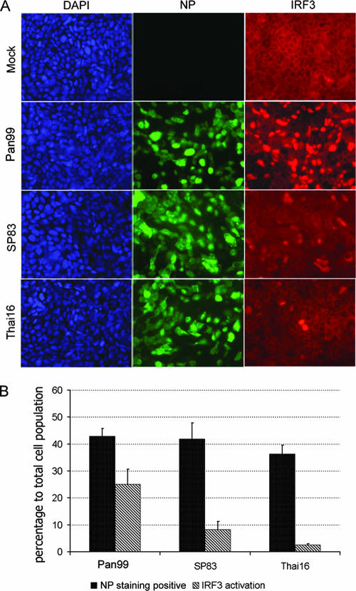
The unparalleled spread of highly pathogenic avian influenza A (HPAI) H5N1 viruses has resulted in devastating outbreaks in domestic poultry and sporadic human infections with a high fatality rate. To better understand the mechanism(s) of H5N1 virus pathogenesis and host responses in humans, we utilized a polarized human bronchial epithelial cell model that expresses both avian alpha-2,3- and human alpha-2,6-linked sialic acid receptors on the apical surface and supports productive replication of both H5N1 and H3N2 viruses. Using this model, we compared the abilities of selected 2004 HPAI H5N1 viruses isolated from humans and a recent human H3N2 virus to trigger the type I interferon (IFN) response. H5N1 viruses elicited significantly less IFN regulatory factor 3 (IRF3) nuclear translocation, as well as delayed and reduced production of IFN-beta compared with the H3N2 virus. Furthermore, phosphorylation of Stat2 and induction of IFN-stimulated genes (ISGs), such as MX1, ISG15, IRF7, and retinoic acid-inducible gene I, were substantially delayed and reduced in cells infected with H5N1 viruses. We also observed that the highly virulent H5N1 virus replicated more efficiently and induced a weaker IFN response than the H5N1 virus that exhibited low virulence in mammals in an earlier study. Our data suggest that the H5N1 viruses tested, especially the virus with the high-pathogenicity phenotype, possess greater capability to attenuate the type I IFN response than the human H3N2 virus. The attenuation of this critical host innate immune defense may contribute to the virulence of H5N1 viruses observed in humans.


Figures

FIG. 1.
FIG. 1.
Polarized human bronchial epithelial Calu-3 cells express both α-2,3- and α-2,6-linked sialic acid receptors. (A and B) Calu-3 cells grown on transwell inserts were fixed and stained for α-2,6-linked sialic acid residues (red) (A) or α-2,3-linked sialic acid (green) (B). The cells were stained with DAPI (blue) to detect cell nuclei. As shown in the insets, treatment with neuraminidase abolished sialic acid residue staining. (C and D) Flow cytometry analysis shows percentages of positive cells for α-2,6 (C)- and α-2,3 (D)-linked sialic acid residues on the Calu-3 cell surface. (E) Immunofluorescence detection of influenza virus NP in Calu-3 cells infected with influenza viruses. Calu-3 monolayers were infected apically with virus at an MOI of 1, stained for NP (red, top panels), and costained with DAPI (blue, bottom panels) at 10 h p.i.
FIG. 2.
FIG. 2.
Transmission electron microscopy demonstrates influenza virus release from the apical surface of polarized Calu-3 cells. Calu-3 cells were infected at the apical surface with virus at an MOI of 5. (A) Mock-infected cells, with the tight junction (arrow), the granular vesicles (arrowhead), and the membrane insert (*) indicated. (B). Thai16 virus-infected cells at 12 h p.i. Both spherical and filamentous virions develop by the positioning of the nucleocapsids at the cell surface (arrowhead). Bars, 1 μm (A) and 100 nm (B).
FIG. 3.
FIG. 3.
Kinetics of influenza virus replication in polarized human bronchial epithelial Calu-3 cells. One-week-old Calu-3 cells grown on transwells were infected apically with the indicated virus at an MOI of 0.01. Culture supernatants were collected at the indicated time points, and viral titers were determined by plaque assay. (A) Replication kinetics of Pan99 virus in Calu-3 cells with or without addition of TPCK-trypsin (0.3 μg/ml). (B) Replication kinetics of three influenza viruses, Pan99, Thai16, and SP83. Asterisks indicate statistically significant differences between SP83 and Thai16 viruses (P < 0.05). (C) Evaluation of the integrity of Calu-3 monolayers after virus infection. Calu-3 monolayers were infected with virus at an MOI of 0.01 and stained with DAPI 48 h later.
FIG. 4.
FIG. 4.
Influenza virus infection results in differential IRF3 nuclear translocation in Calu-3 cells. Polarized Calu-3 cells were infected with viruses at an MOI of 1. Cells were fixed and stained for influenza virus NP (green) and IRF3 (red) at 8 h p.i. (A) Immunofluorescent staining of virus-infected cells. (B) Assessment of rate of infection and IRF3 nuclear translocation in virus-infected cells. Error bars indicate standard deviations.
FIG. 5.
FIG. 5.
Primary IFN response to influenza virus infection in Calu-3 cells. (A) Evaluation of IFN-β mRNA expression by RT-PCR. Total RNA was isolated from Calu-3 cells infected with virus at an MOI of 1 or 3 for 8 h. The data are presented as the relative fold gene induction and are representative of three independent replicates. (B) Assessment of activation of the JAK-STAT pathway induced by virus infection. Protein samples from the indicated treatments were harvested and examined by Western blotting for STAT2 phosphorylation. C, control.
FIG. 6.
FIG. 6.
Transcription of the IFN-β gene in NHBE cells in response to influenza virus infection. Confluent NHBE cells grown on inserts were infected with virus at an MOI of 5. (A) Immunofluorescence detection of influenza virus NP protein in infected cells. (B) Examination of IFN-β expression by real-time RT-PCR.
FIG. 7.
FIG. 7.
Production of IFN-β in influenza virus-infected Calu-3 cells. Polarized Calu-3 cells were infected with virus at an MOI of 1. (A) Evaluation of IFN-β production by ELISA. Supernatants from infected Calu-3 cells, collected at the designated times, were analyzed by ELISA. Three sets of samples from independent treatments were tested in duplicate. Single asterisks indicate statistically significant differences between human H3N2 virus and avian viruses (P < 0.05), and double asterisks indicate statistically significant differences between two avian viruses (P < 0.05). Error bars indicate standard deviations. (B) Analysis of IFN-β expression in virus-infected Calu-3 cells by RT-PCR. The data are presented as the relative fold gene induction and are representative of three independent replicates. (C) Activation of the JAK-STAT pathway during influenza virus infection. Phosphorylation of STAT2 is shown in the top panel and β-actin in the bottom panel in the Western blot. C, control.
FIG. 8.
FIG. 8.
Expression of ISGs in infected Calu-3 cells. Polarized Calu-3 cells were infected with virus at an MOI of 1, and total RNA was isolated and examined with real-time PCR. Expression analysis of four genes, encoding myxovirus (influenza virus) resistance 1 (Mx1), IFN-induced protein IFI-15K (ISG15), RIG-I, and IRF7, is shown. Expression of the viral M1 gene also was determined; at 8 h p.i. expression of M1 in SP83-infected cells was lower than that for the Thai16 and Pan99 viruses (data not shown). The data are presented as the relative fold gene induction and are representative of three independent replicates.
FIG. 9.
FIG. 9.
Pretreatment with IFN-β results in delay and reduction of viral replication. Calu-3 cells were pretreated with 200 U/ml of human IFN-β (Sigma, St. Louis, MO) for 24 h and were then infected with viruses at an MOI of 0.01. Supernatants were collected at the indicated time points, and virus titers were measured by plaque assay. Error bars indicate standard deviations.

References

    1. Chan, M. C., C. Y. Cheung, W. H. Chui, S. W. Tsao, J. M. Nicholls, Y. O. Chan, R. W. Chan, H. T. Long, L. L. Poon, Y. Guan, and J. S. Peiris. 2005. Proinflammatory cytokine responses induced by influenza A (H5N1) viruses in primary human alveolar and bronchial epithelial cells. Respir. Res. 6:135. - PMC - PubMed
    1. Chotpitayasunondh, T., K. Ungchusak, W. Hanshaoworakul, S. Chunsuthiwat, P. Sawanpanyalert, R. Kijphati, S. Lochindarat, P. Srisan, P. Suwan, Y. Osotthanakorn, T. Anantasetagoon, S. Kanjanawasri, S. Tanupattarachai, J. Weerakul, R. Chaiwirattana, M. Maneerattanaporn, R. Poolsavathitikool, K. Chokephaibulkit, A. Apisarnthanarak, and S. F. Dowell. 2005. Human disease from influenza A (H5N1), Thailand, 2004. Emerg. Infect. Dis. 11:201-209. - PMC - PubMed
    1. Coro, E. S., W. L. Chang, and N. Baumgarth. 2006. Type I IFN receptor signals directly stimulate local B cells early following influenza virus infection. J. Immunol. 176:4343-4351. - PubMed
    1. Davies, J. R., S. Kirkham, N. Svitacheva, D. J. Thornton, and I. Carlstedt. 2007. MUC16 is produced in tracheal surface epithelium and submucosal glands and is present in secretions from normal human airway and cultured bronchial epithelial cells. Int. J. Biochem. Cell. Biol. 39:1943-1954. - PubMed
    1. de Jong, M. D., V. C. Bach, T. Q. Phan, M. H. Vo, T. T. Tran, B. H. Nguyen, M. Beld, T. P. Le, H. K. Truong, V. V. Nguyen, T. H. Tran, Q. H. Do, and J. Farrar. 2005. Fatal avian influenza A (H5N1) in a child presenting with diarrhea followed by coma. N. Engl. J. Med. 352:686-691. - PubMed

MeSH terms