. 2007 Nov;81(22):12427-38.
doi: 10.1128/JVI.01105-07. Epub 2007 Sep 12.

Influenza virus mRNA translation revisited: is the eIF4E cap-binding factor required for viral mRNA translation?

Affiliations

Influenza virus mRNA translation revisited: is the eIF4E cap-binding factor required for viral mRNA translation?

Idoia Burgui et al. J Virol. 2007 Nov.

Abstract

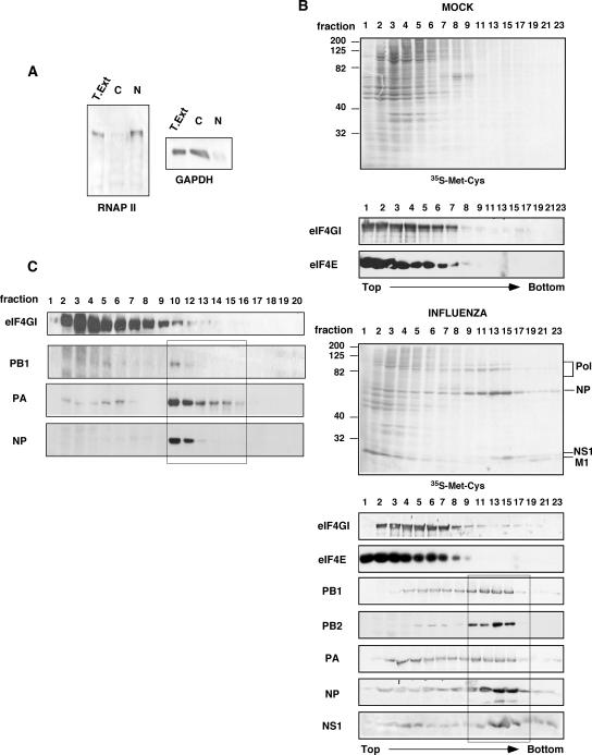
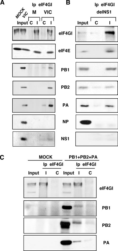
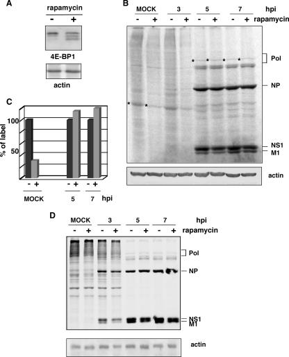
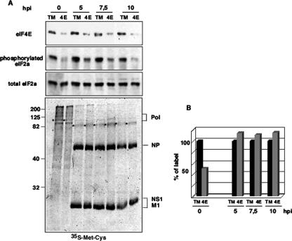
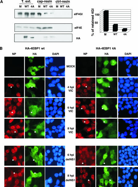
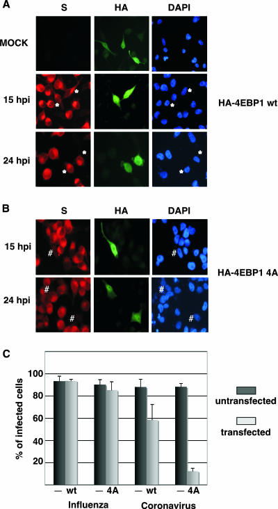
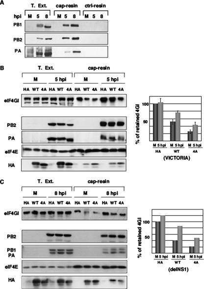
Influenza virus mRNAs bear a short capped oligonucleotide sequence at their 5' ends derived from the host cell pre-mRNAs by a "cap-snatching" mechanism, followed immediately by a common viral sequence. At their 3' ends, they contain a poly(A) tail. Although cellular and viral mRNAs are structurally similar, influenza virus promotes the selective translation of its mRNAs despite the inhibition of host cell protein synthesis. The viral polymerase performs the cap snatching and binds selectively to the 5' common viral sequence. As viral mRNAs are recognized by their own cap-binding complex, we tested whether viral mRNA translation occurs without the contribution of the eIF4E protein, the cellular factor required for cap-dependent translation. Here, we show that influenza virus infection proceeds normally in different situations of functional impairment of the eIF4E factor. In addition, influenza virus polymerase binds to translation preinitiation complexes, and furthermore, under conditions of decreased eIF4GI association to cap structures, an increase in eIF4GI binding to these structures was found upon influenza virus infection. This is the first report providing evidence that influenza virus mRNA translation proceeds independently of a fully active translation initiation factor (eIF4E). The data reported are in agreement with a role of viral polymerase as a substitute for the eIF4E factor for viral mRNA translation.


Figures

FIG. 1.
FIG. 1.
Separation of influenza virus polymerase from viral RNPs. HEK293T cells were mock or influenza virus infected for 7 h with the VIC strain at 5 to 10 PFU/cell. (A) Subcellular fractionation. Nuclear (N) and cytosolic (C) fractions from total extracts (T. Ext.) were separated and analyzed by Western blotting with specific antibodies against RNA polymerase II (RNAP II) or GAPDH. (B) Separation of viral polymerase subunits from viral RNPs. HEK293T cells were labeled in vivo during the last 4 h of infection (Promix; Amersham). Next, the cells were collected and processed as described in Materials and Methods, and the labeled proteins were analyzed by autoradiography ([35S]Met-Cys). Samples of the same fractions were analyzed by Western blotting with specific antibodies against the indicated proteins. (C) Viral RNP expressing NS1 was reconstituted in vivo and processed as described above (B). The corresponding proteins were analyzed by Western blotting.
FIG. 2.
FIG. 2.
Influenza virus polymerase subunits associate with translation initiation complexes. (A) Cytosolic extracts from mock-infected or VIC-infected HEK293T cells were applied to sucrose gradients and processed as described in Materials and Methods. Samples were used for immunoprecipitation studies (Ip) using specific antibodies against the eIF4GI protein (I) or the preimmune serum (C). (B) HEK293T cells were infected with the delNS1 strain, and cytosolic extracts were immunoprecipitated as described above (A) to analyze the associated proteins by Western blotting. (C) HEK293T cells were untransfected (MOCK) or cotransfected with plasmids expressing PB1, PB2, and PA (PB1+PB2+PA), and cytosolic extracts were prepared and immunoprecipitated with eIF4GI antiserum as described above. The polymerase proteins associated with eIF4GI were analyzed by Western blotting.
FIG. 3.
FIG. 3.
Influenza virus infection progresses efficiently in rapamycin-treated cells. (A) HeLa cells treated with (+) or without (−) rapamycin for 12 h were subjected to Western blotting against 4E-BP1 or actin. (B) HeLa cells treated with (+) or without (−) rapamycin for 12 h were mock infected (MOCK) or infected with the VIC strain, maintaining the rapamycin condition. At the indicated hpi, the cells were metabolically labeled, and the proteins were analyzed by SDS-polyacrylamide gels and autoradiography. (C) Quantitation of the incorporated label in specific cellular and viral proteins (marked with asterisks) from B. (D) A549 cells treated with (+) or without (−) rapamycin were processed as indicated above (B).
FIG. 4.
FIG. 4.
Gene silencing of the eIF4E factor does not affect influenza virus protein synthesis but inhibits cellular protein translation. HEK293T cells were transfected with control pSUPER-GFP-TM (TM) or pSUPER-GFP-4E (4E) plasmids. Twelve hours after transfection, the cells were selected by cell sorting using the GFP fluorescence and plated again. Thirty six hours posttransfection, cells were infected with influenza virus. (A) At the indicated hpi, aliquots were taken and used for Western blotting against the indicated proteins and metabolic labeling with [35S]Met-Cys. (B) Quantitation of the incorporated label in specific cellular and viral proteins (marked with asterisks) from the [35S]Met-Cys panel.
FIG. 5.
FIG. 5.
Overexpression of HA-4E-BP1 proteins does not affect influenza virus infection in HEK293T cells. (A) HEK293T cells were untransfected (M) or transfected with plasmids expressing HA-tagged wild-type 4E-BP1 (WT) or nonphosphorylatable 4E-BP1 (4A) protein. At 36 h posttransfection, total cell extracts (T.ext.) were used to study eIF4GI, eIF4E, and HA-4E-BP1 (HA) retention either to cap resins or to Sepharose-4B control resins (ctrl-resin) by Western blotting. Quantitation of eIF4GI protein retained on the cap resins is shown on the right. (B) HEK293T cells were transfected with plasmid pcDNA3-HA-4E-BP1 wt or pcDNA3-HA-4E-BP1 4A, and 36 h posttransfection, the cells were mock or influenza virus infected with either the VIC (top) or delNS1 (bottom) strain. At the indicated hpi, cells were fixed and used for immunofluorescence using antibodies against HA to monitor plasmid transfection and NP protein to monitor influenza virus infection. Asterisks indicate transfected and infected cells. DAPI, 4′,6′-diamidino-2-phenylindole.
FIG. 6.
FIG. 6.
Overexpression of underphosphorylated 4E-BP1 protein impairs human coronavirus infection. HeLa cells were transfected with plasmid pcDNA3-HA-4E-BP1 wt (A) or pcDNA3-HA-4E-BP1 4A (B), and 36 h posttransfection, the cells were mock or coronavirus infected. At the indicated hpi, cells were used for immunofluorescence using antibodies against HA to monitor plasmid transfection and S protein to monitor coronavirus infection. Asterisks indicate transfected and infected cells. # indicates cells that were transfected but uninfected. DAPI, 4′,6′-diamidino-2-phenylindole. (C) Quantitation of the efficiency of infection in untransfected cells (−) and in pcDNA3-HA-4E-BP1 wt (WT)- or pcDNA3-HA-4E-BP1 4A (4A)-transfected cells subsequently infected with influenza virus (data not shown) or human coronavirus HCoV-229E.
FIG. 7.
FIG. 7.
Influenza virus infection enhances the recruitment of eIF4GI to cap resins. (A) Cytosolic extracts (T. Ext.) of mock-infected (M) or influenza virus-infected (5 and 8 hpi) HEK293T cells were applied to m7GTP-Sepharose resins (cap resins) or to Sepharose-4B control resins (ctrl-resins), and the indicated proteins were analyzed by Western blotting. (B) HEK293T cells were transfected with plasmid pcDNA3-HA (HA), pcDNA3-HA-4E-BP1 wt (WT), or pcDNA3-HA-4E-BP1 4A (4A), and 36 h posttransfection, the cells were mock (M) or influenza virus infected (5 hpi). Cytosolic extracts (T. Ext.) were prepared and applied to m7GTP-Sepharose resins (cap-resin). Retention of the indicated proteins was evaluated by Western blot analysis, and quantitation is shown on the right (means and standard deviations). (C) Experiment similar to that performed in B but using recombinant delNS1 influenza virus. On the right side, the quantitation of the eIF4GI protein retained in the cap resins as described in the text is shown.

References

    1. Aragón, T., S. de la Luna, I. Novoa, L. Carrasco, J. Ortín, and A. Nieto. 2000. Eukaryotic translation initiation factor 4GI is a cellular target for NS1 protein, a translational activator of influenza virus. Mol. Cell. Biol. 20:6259-6268. - PMC - PubMed
    1. Beloso, A., C. Martínez, J. Valcárcel, J. Fernández-Santarén, and J. Ortín. 1992. Degradation of cellular mRNA during influenza virus infection: its possible role in protein synthesis shutoff. J. Gen. Virol. 73:575-581. - PubMed
    1. Beretta, L., A. C. Gingras, Y. V. Svitkin, M. N. Hall, and N. Sonenberg. 1996. Rapamycin blocks the phosphorylation of 4E-BP1 and inhibits cap-dependent initiation of translation. EMBO J. 15:658-664. - PMC - PubMed
    1. Blaas, D., E. Patzelt, and E. Keuchler. 1982. Identification of the cap binding protein of influenza virus. Nucleic Acids Res. 10:4803-4812. - PMC - PubMed
    1. Burgui, I., T. Aragón, J. Ortín, and A. Nieto. 2003. PABP1 and eIF4GI associate to influenza virus NS1 protein in viral mRNA translation initiation complexes. J. Virol. 84:3263-3274. - PubMed

Publication types

MeSH terms