Comparative Study
. 2007 Sep 12;27(37):9962-74.
doi: 10.1523/JNEUROSCI.1536-07.2007.

Properties of persistent postnatal cortical subplate neurons

Affiliations
Comparative Study

Properties of persistent postnatal cortical subplate neurons

Juan Torres-Reveron et al. J Neurosci. .

Abstract

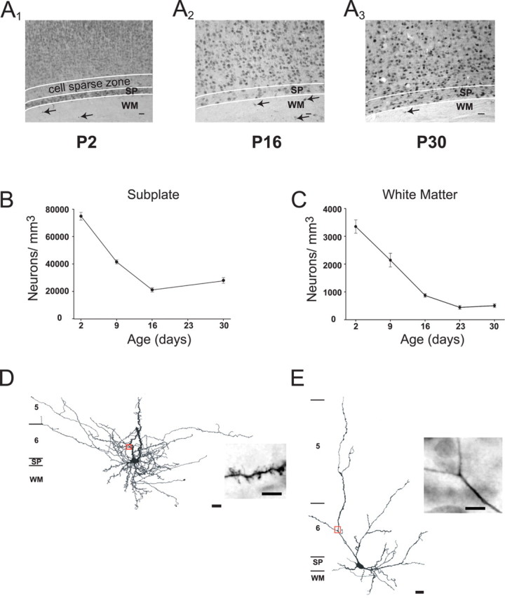
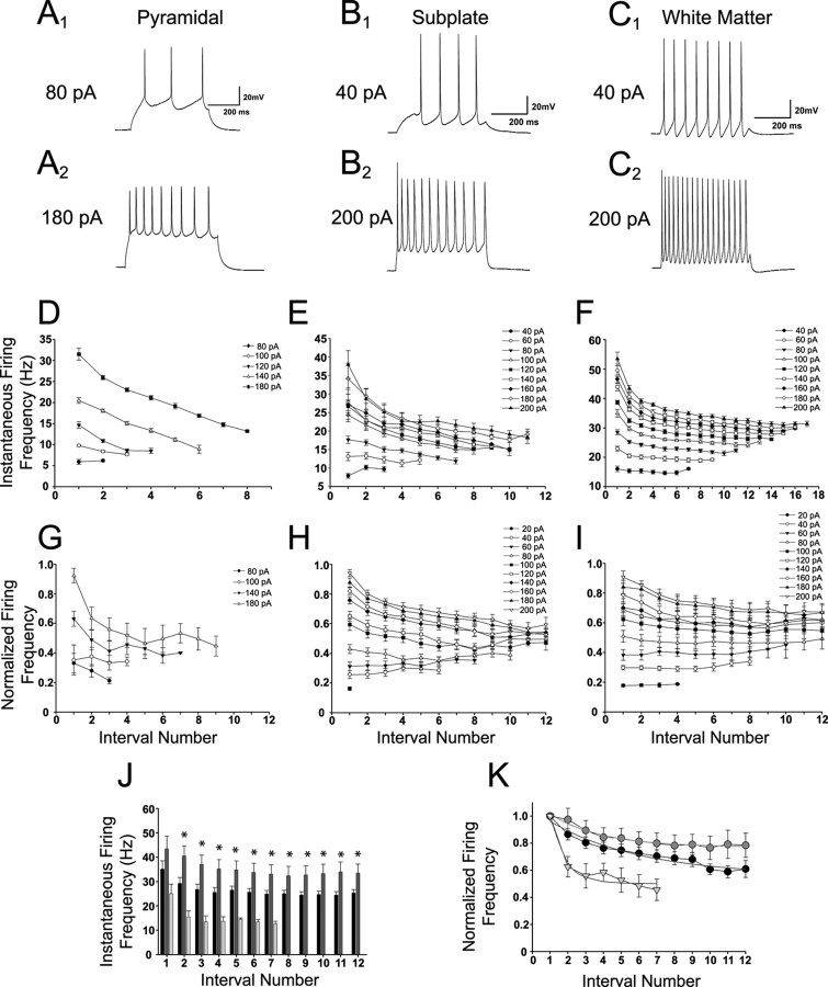
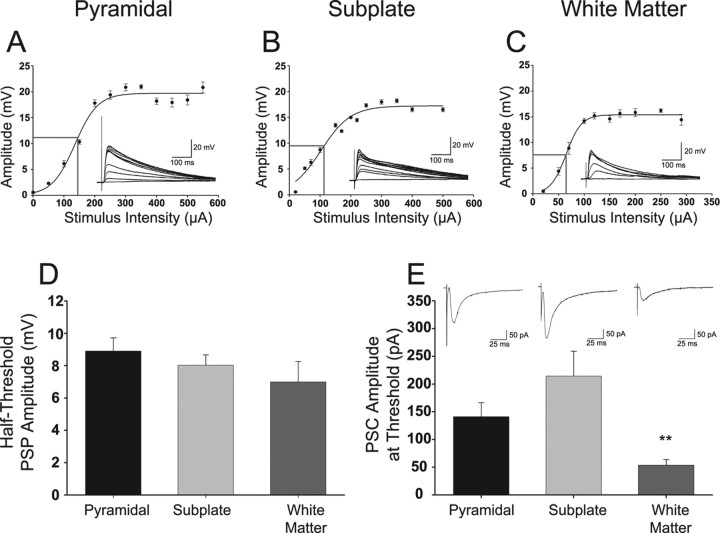
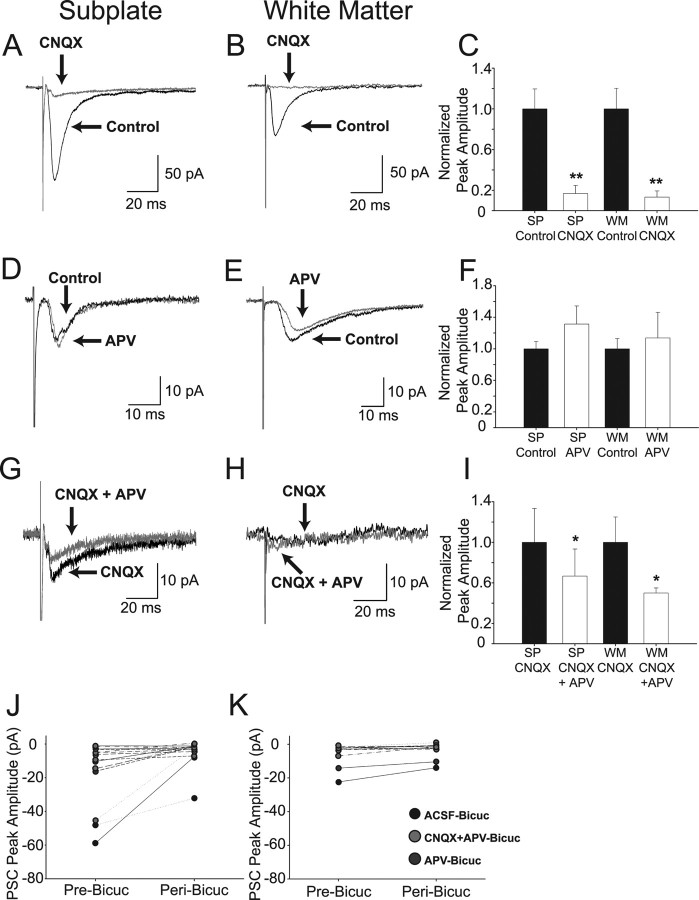
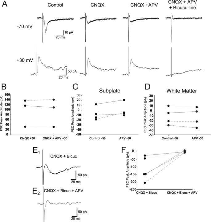
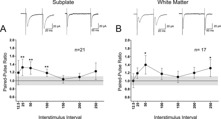
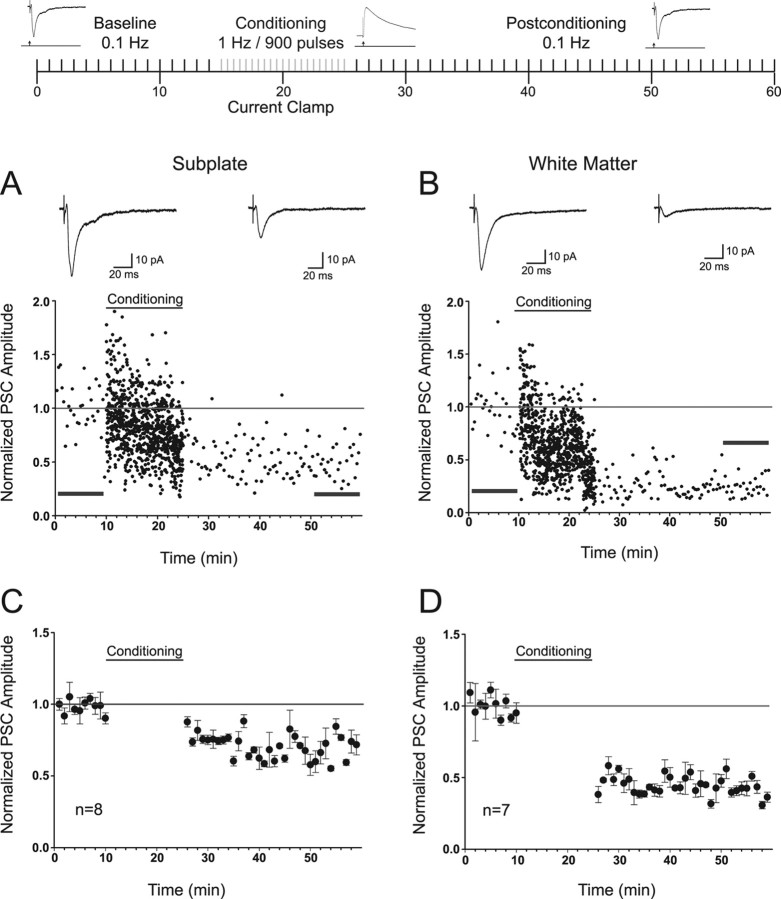
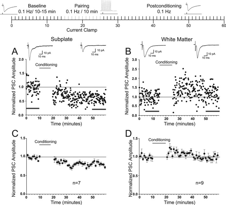
Subplate (SP) neurons are important for the proper development of thalamocortical innervation. They are necessary for formation of ocular dominance and orientation columns in visual cortex. During the perinatal period, many SP neurons die. The surviving cohort forms interstitial cells in the white matter (WM) and a band of horizontally oriented cells below layer VI (layer VIb, layer VII, or subplate cells). Although the function of embryonic SP neurons has been well established, the functional roles of WM and postnatal SP cells are not known. We used a combination of anatomical, immunohistochemical, and electrophysiological techniques to explore the dendritic morphology, neurotransmitter phenotype, intrinsic electrophysiological, and synaptic input properties of these surviving cells in the rat visual cortex. The density of SP and WM cells significantly decreases during the first month of life. Both populations express neuronal markers and have extensive dendritic arborizations within the SP, WM, and to the overlying visual cortex. Some intrinsic electrophysiological properties of SP and WM cells are similar: each generates high-frequency slowly adapting trains of action potentials in response to a sustained depolarization. However, SP cells exhibit greater frequency-dependent action potential broadening than WM neurons. Both cell types receive predominantly AMPA/kainate receptor-mediated excitatory synaptic input that undergoes paired-pulse facilitation as well as NMDA receptor and GABAergic input. Synaptic inputs to these cells can also undergo long-term synaptic plasticity. Thus, surviving SP and WM cells are functional electrogenic neurons integrated within the postnatal visual cortical circuit.


Figures

Figure 1.
Figure 1.
SP and WM cells decrease in density during the first month postnatally. A, Representative examples of NeuN-stained rat visual cortex at P2 (A1), P16 (A2), and P30 (A3). Filled arrows indicate WM cells at each age. B, C, Plots of neuronal density of SP (B) and WM (C) cells during the first month after birth in rat visual cortex. A developmental decrease was observed in both populations. D, E, Camera lucida drawings of a spiny (D) and smooth (E) SP cell. Insets are photomicrographs of the area of dendrite indicated by the red box in each drawing. Approximate location of the layers is indicated on the left of each drawing. Scale bars: photomicrographs, 50 μm; drawings, 20 μm; insets, 5 μm.
Figure 2.
Figure 2.
SP and WM cells exhibit slow spike frequency adaptation. Traces and graphs shown for each group are arranged in columns. A–C, Representative traces for a pyramidal (A1, A2), SP (B1, B2), and WM (C1, C2) cell at the depolarization level indicated on the left. D–F, Instantaneous spike frequency analysis for the cell shown in the examples for pyramidal (D), SP (E), and WM (F). Each line represents a different depolarization level, and each point is a particular interval number. G–I, Analysis of spike frequency adaptation group data in pyramidal (G), SP (H), and WM (I) cells. Data were normalized to the largest instantaneous current in each cell. Traces shown for pyramidal cells start at a higher depolarization level because spikes were rarely present below 80 pA. J, Comparison of instantaneous frequency for each cell group versus spike interval number at 200 pA depolarization. K, Comparison of the adaptation rate between each cell group. Gray lines are single-exponential fits. For J and K, pyramidal cells are indicated by light gray bars and triangles, SP cells are indicated by black bars and circles, and WM cells are indicated by dark gray bars and gray circles. Significance calculated for the difference between SP and WM neurons. *p < 0.05.
Figure 3.
Figure 3.
Synaptic input–output properties in pyramidal, SP, and WM cells. A–C, Representative examples of input–output curves in pyramidal (A), SP (B), and WM (C) cells. Gray lines indicate the half-threshold PSP amplitude and stimulus intensity for each cell. Insets are PSPs for each indicated cell type on top. D, Plot of average ± SEM half-threshold PSP amplitude in pyramidal (black bar; n = 6), SP (light gray bar; n = 6) and WM (dark gray bar; n = 4) neurons. E, Plot of the average ± SEM. PSC amplitude at threshold evoked in voltage clamp at a synaptic stimulating current amplitude that was used for that same cell in current-clamp mode to evoke a PSP that was just below spike threshold for pyramidal (black bar; n = 6), SP (light gray bar; n = 6), and WM (dark gray bar; n = 10) cells. Insets are PSCs for each cell type below. Statistical comparisons are between WM and the other cell groups. **p < 0.01.
Figure 4.
Figure 4.
Effect of AMPA/kainate, NMDA, and GABAA receptor pharmacological inhibition on synaptic responses evoked in SP and WM cells. Panels correspond to the cell type (SP or WM) indicated at the top of each column. AC, Effects of CNQX alone on synaptic responses evoked at −70 mV. Representative examples of averaged evoked synaptic responses from an SP (A) and a WM (B) cell in aCSF (control; large black traces) and in CNQX (small gray traces). Summary of effects of CNQX on normalized peak evoked synaptic responses at −70 mV (C). D–F, Effect of APV alone on evoked synaptic currents at −70 mV. Representative examples of averaged evoked synaptic responses from an SP (D) and a WM (E) cell in aCSF (control; black traces) and in APV (gray traces). Summary of effects of APV on normalized peak evoked synaptic currents at −70 mV (F). G–I, Effect of APV and CNQX on evoked synaptic currents at −70 mV. Small currents remain in some cells at −70 mV after blockade of AMPA/kainate receptors with CNQX. Representative examples of average evoked synaptic responses from an SP (G) and a WM (H) cell in CNQX alone (black traces) and in CNQX/APV (gray traces). Summary of effect of APV/CNQX (vs CNQX alone) on normalized peak evoked synaptic responses at −70 mV (I). J, K, Effects of bicuculline on amplitude of average peak evoked synaptic responses of SP (J) and WM (K) cells. The peak responses are plotted for each individual cell tested in the control (Pre-Bicuc) condition and the experimental (Peri-Bicuc) condition. The control responses varied in their amplitude ranges as the bicuculline was applied after a no-drug control condition (aCSF), in conjunction with APV after application of APV alone, or in conjunction with CNQX/APV after application of CNQX/APV. *p < 0.05; **p < 0.01.
Figure 5.
Figure 5.
Effects of APV application on evoked synaptic responses in the presence of CNQX at −70 and +30 mV before and after application of bicuculline. Example (A) synaptic responses of an SP cell recorded at −70 and +30 mV holding potential. An inward (control, −70 mV) and the corresponding outward (+30 mV) current are seen at the two holding potentials. The current is significantly decreased by CNQX at −70 mV (−52%; p < 0.01) but is not significantly affected by APV (−3%; p > 0.05) at either potential. Bicuculline eliminated the remaining current at both membrane potentials. Individual (B) data for SP cells (n = 3) that were depolarized to +30 mV in the presence of CNQX (CNQX + 30) and in the presence of CNQX/APV (CNQX + APV + 30). The NMDA receptor component was not observed when SP (n = 4) (C) or WM (n = 4) (D) cells were depolarized to −50 mV. Inward PSCs are seen at +30 mV when SP cells are recorded in low Mg2+/high Ca2+ and during blockade of AMPA/kainate and GABAA receptors (E1). The current is eliminated by application of APV (E2). Individual data (F) for SP cells (n = 5) depolarized to +30 mV in the presence of CNQX and bicuculline (CNQX + Bicuc) and in the presence of CNQX, bicuculline, and APV (CNQX + Bicuc + APV).
Figure 6.
Figure 6.
Responses of SP and WM cells to paired-pulse synaptic stimulation. Inset traces at top are recorded averaged paired-pulse evoked PSCs from an example SP (left) and WM (right) cell for the 50 and 200 ms ISI. A, B, Analysis of group PPR in SP (A; n = 21) and WM (B; n = 17) cells. Horizontal lines at 1.0 PPR indicate the interface between paired-pulse depression (<1.0) and paired-pulse facilitation (>1.0). Gray region indicates the variability in the PPR expected from normal variability of a single PSC (the mean ± SEM PPR for the ratio of the first PSC in each trial related to the first PSC in the previous trial with 5 s intertrial intervals). Filled circles in the group data represent the mean ± SEM of the PPR at each ISI. *p < 0.05; **p < 0.01.
Figure 7.
Figure 7.
Responses of SP and WM cells to 1 Hz synaptic conditioning for plasticity induction. Schematic shows the organization of the experimental protocol (for details, see Materials and Methods). Inset traces are examples of the recorded PSCs and PSPs in current and voltage clamp, respectively. Arrow in the insets represents the location of extracellular stimulation. Gray bars indicate the conditioning period. A, B, Representative time plots of PSCs before and after 15 min 1 Hz synaptic stimulation for an individual SP and WM cell that exhibited LTD, respectively. Insets are average PSCs 10 min before 1 Hz stimulation and from 25–35 min after 1 Hz stimulation (horizontal bars). Horizontal line indicates the interface between depression and facilitation. C, D, Group data for all SP (n = 8) and WM (n = 7) cells, respectively. Significant LTD (p < 0.001) was observed in both cases. The mean group LTD in SP cells was 31%, whereas it was 55% in WM cells.
Figure 8.
Figure 8.
Responses of SP and WM cells to plasticity induction protocol that pairs synaptic input at 0.1 Hz with direct depolarization (and spiking) of the postsynaptic cell. Schematic shows the organization of the pairing protocol used for LTP induction (for details, see Materials and Methods). Inset traces are examples of whether the recording was in voltage or current clamp and are not meant to represent a particular experiment. Arrow in the insets represents the location of extracellular stimulation. Gray bars indicate the conditioning period. Spike train represents the depolarization of the cell during the pairing. Representative examples from SP (A), showing significant depression, and WM (B), showing significant potentiation. Insets are average PSCs 10 min before pairing and from 30–40 min after conditioning (horizontal bars). C, D, Group data for all SP (n = 7) and WM (n = 9) cells in the sample, respectively. Significant depression (19%) was seen in SP cells (p < 0.001), but no change (3%) was seen in WM cells because some cells also showed mild potentiation.

References

    1. Abraham WC, Bear MF. Metaplasticity: the plasticity of synaptic plasticity. Trends Neurosci. 1996;19:126–130. - PubMed
    1. Aggoun-Aouaoui D, Kiper DC, Innocenti GM. Growth of callosal terminal arbors in primary visual areas of the cat. Eur J Neurosci. 1996;8:1132–1148. - PubMed
    1. Al Ghoul WM, Miller MW. Transient expression of Alz-50 immunoreactivity in developing rat neocortex: a marker for naturally occurring neuronal death? Brain Res. 1989;481:361–367. - PubMed
    1. Aldrich RW, Jr, Getting PA, Thompson SH. Mechanism of frequency-dependent broadening of molluscan neurone soma spikes. J Physiol (Lond) 1979;291:531–544. - PMC - PubMed
    1. Allendoerfer KL, Shatz CJ. The subplate, a transient neocortical structure: its role in the development of connections between thalamus and cortex. Annu Rev Neurosci. 1994;17:185–218. - PubMed

Publication types

LinkOut - more resources