Comparative Study
. 2007 Sep 12;27(37):10007-14.
doi: 10.1523/JNEUROSCI.2862-07.2007.

The itch-producing agents histamine and cowhage activate separate populations of primate spinothalamic tract neurons

Affiliations
Comparative Study

The itch-producing agents histamine and cowhage activate separate populations of primate spinothalamic tract neurons

Steve Davidson et al. J Neurosci. .

Abstract

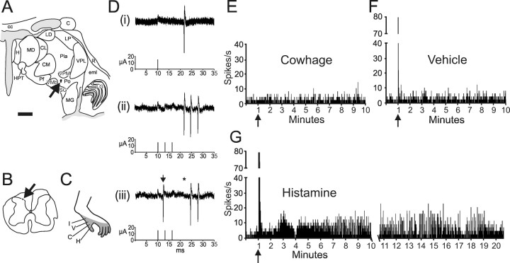
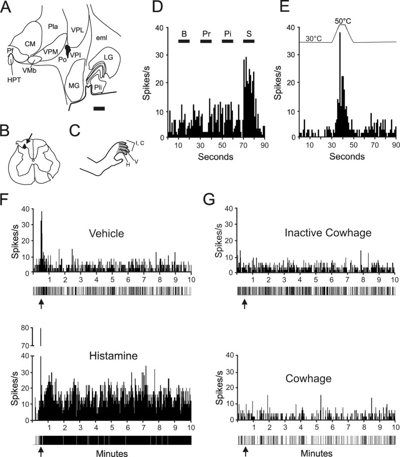
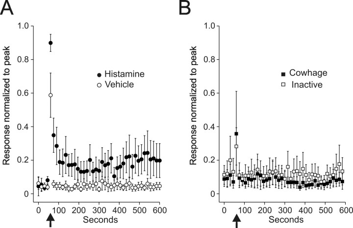
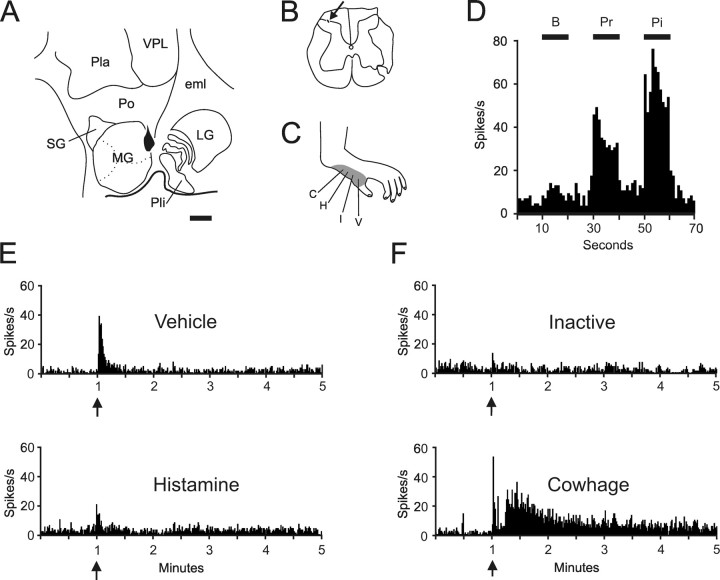
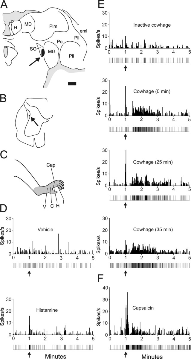
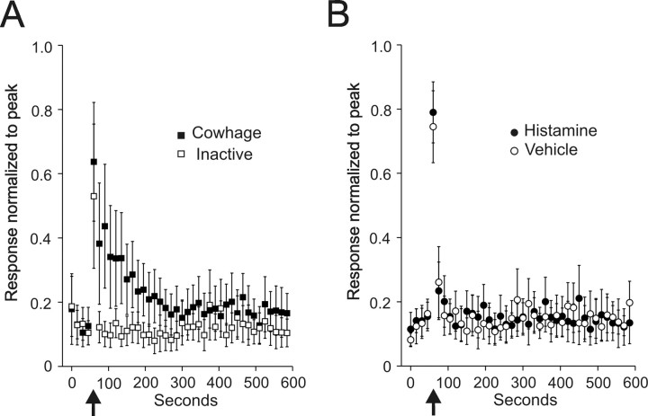
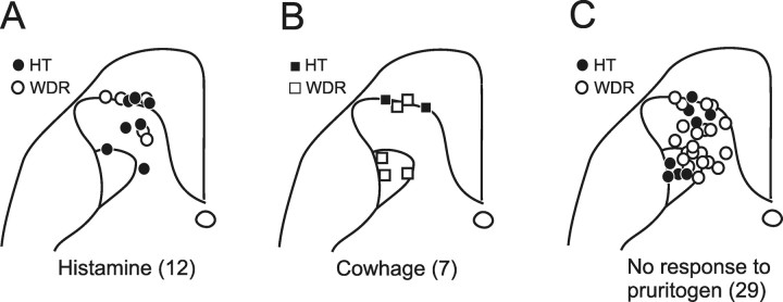
Itch is an everyday sensation, but when associated with disease or infection it can be chronic and debilitating. Several forms of itch can be blocked using antihistamines, but others cannot and these constitute an important clinical problem. Little information is available on the mechanisms underlying itch that is produced by nonhistaminergic mechanisms. We examined the responses of spinothalamic tract neurons to histaminergic and, for the first time, nonhistaminergic forms of itch stimuli. Fifty-seven primate spinothalamic tract (STT) neurons were identified using antidromic activation techniques and examined for their responses to histamine and cowhage, the nonhistaminergic itch-producing spicules covering the pod of the legume Mucuna pruriens. Each examined neuron had a receptive field on the hairy skin of the hindlimb and responded to noxious mechanical stimulation. STT neurons were tested with both pruritogens applied in a random order and we found 12 that responded to histamine and seven to cowhage. Each pruritogen-responsive STT neuron was activated by the chemical algogen capsaicin and two-thirds responded to noxious heat stimuli, demonstrating that these neurons convey chemical, thermal, and mechanical nociceptive information as well. Histamine or cowhage responsive STT neurons were found in both the marginal zone and the deep dorsal horn and were classified as high threshold and wide dynamic range. Unexpectedly, histamine and cowhage never activated the same cell. Our results demonstrate that the spinothalamic tract contains mutually exclusive populations of neurons responsive to histamine or the nonhistaminergic itch-producing agent cowhage.


Figures

Figure 1.
Figure 1.
Antidromic activation of a histamine-responsive STT neuron. A, Location of the lesion at the stimulating point in the posterior nucleus. Scale bar, 2.0 mm. C, Caudate; cc, corpus callosum; CL, center lateral nucleus (n.); CM, center median n.; eml, external medullary lamina; H, habenula; HPT, habenulopeduncular tract; LD, lateral dorsal n.; LG, lateral geniculate n.; LP, lateral posterior n.; MD, mediodorsal n.; MG, medial geniculate n.; Pf, parafascicular n.; Pla, anterior pulvinar; Pli, inferior pulvinar; Pll, lateral pulvinar; Plm, medial pulvinar; Po, posterior n; R, reticular n; SG, suprageniculate n.; VMb, ventrobasal part of the ventral medial n.; VPI, ventral posterior inferior n.; VPM, ventral posterior medial n. B, Location of the recording point in the marginal zone of lumbar spinal cord (arrow). C, Receptive field. Letters indicate the location of each stimulus. H, Histamine; V, vehicle; C, cowhage; I, inactive cowhage. D, Five overlaid traces demonstrate the constant latency of 11.6 ms from the stimulation (15 μA) to recording (i). The neuron followed a >300 Hz train without failure (ii), and an ascending orthodromic action potential (iii, arrow) collided with an antidromic action potential (asterisk located at the anticipated latency). E, F, Stimuli were applied in the order illustrated. This neuron was not excited by cowhage or vehicle applied to the receptive field by a cotton tipped swab. Bin width for this and all single unit histograms is 1 s. G, The response to histamine lasted 20 min.
Figure 2.
Figure 2.
Example of a HT histamine responsive spinothalamic tract neuron. A, Location of the lesion marking the stimulating point in the ventral posterior inferior nucleus of the thalamus. The axon was antidromically activated with 27 μA. Scale bar, 1.0 mm. B, Lesion at the recording point in the marginal zone. C, The receptive field extended over the hairy skin of four toes. D, Histogram of responses during brush (B), pressure (Pr), pinch (Pi), and squeeze (S) of the receptive field. E, Response of this neuron to a heat ramp to 50°C. F, Vehicle (top) and histamine (bottom) were injected intradermally (arrows). Impulse record is shown below the histogram. G, Inactive cowhage (top) and cowhage (bottom) were applied (arrows) to the skin via an M. pruriens pod. See Figure 1 legend for abbreviations.
Figure 3.
Figure 3.
Mean ± SEM of 12 histamine responsive STT units normalized to the peak discharge during intradermal histamine injection. Data points are the mean of consecutive 15 s bins. A, Response to histamine and vehicle. Arrows indicate the time of injection. In some cells, the largest number of action potentials per 15 s bin came after the injection; therefore, the normalized peak did not equal 1.0. B, Response of the same cells in A to inactive and active cowhage.
Figure 4.
Figure 4.
Example of a cowhage responsive spinothalamic tract neuron. A, Location of the lesion marking the stimulating point on the border of the medial geniculate nucleus and the posterior nucleus of the thalamus. The antidromic threshold was 25 μA. Scale bar, 1.0 mm. B, Recording point in the marginal zone. C, Receptive field over the hairy skin of hind paw. D, Histogram of responses to mechanical stimulation. This unit was classified WDR. E, Vehicle and histamine produced a brief discharge during the intradermal injection. F, Inactive cowhage produced no discharge, but cowhage produced a response lasting ∼4 min after a latent period. See Figure 1 legend for abbreviations.
Figure 5.
Figure 5.
Example of the repeatability of the response to cowhage. A, Location of antidromic stimulating point (24 μA) in the suprageniculate n. of the thalamus. Scale bar, 1.0 mm. B, Recording point in the lateral reticulated area. C, Receptive field over the entire hind paw. Cap, Capsaicin. D, Intradermal injection of vehicle and histamine. Impulse record is shown below the histogram. E, Application of inactive cowhage (top) and cowhage at three time points. F, Response to 10 μg of capsaicin. See Figure 1 legend for abbreviations.
Figure 6.
Figure 6.
Mean ± SEM of seven cowhage-responsive STT units normalized to the peak discharge during intradermal injection of histamine. A, Inactive and active cowhage. B, Responses from the same cells to vehicle and histamine.
Figure 7.
Figure 7.
Location in the dorsal horn of each pruritogen responsive STT neuron with HT or WDR classification indicated. A, Histamine responsive STT neurons were mostly located in the marginal zone and the nucleus proprius. Cowhage responsive units were located within the marginal zone and also within the lateral reticulated area. C, Location of the 29 recovered recording points of the 38 total STT units not excited by either histamine or cowhage.

References

    1. Andrew D, Craig AD. Spinothalamic lamina I neurons selectively sensitive to histamine: a central neural pathway for itch. Nat Neurosci. 2001;4:72–77. - PubMed
    1. Bickford RG. Experiments relating to the itch sensation, its peripheral mechanism and central pathways. Clin Sci. 1938;3:377–386.
    1. Burstein R, Dado RJ, Cliffer KD, Giesler GJ., Jr Physiological characterization of spinohypothalamic tract neurons in the lumbar enlargement of rats. J Neurophysiol. 1991;66:261–284. - PubMed
    1. Carstens E. Responses of rat spinal dorsal horn neurons to intracutaneous injection of histamine, capsaicin and other irritants. J Neurophysiol. 1997;77:2499–2514. - PubMed
    1. Dado RJ, Katter JT, Giesler GJ., Jr Spinothalamic and spinohypothalamic tract neurons in the cervical enlargement of rats. I. Locations of antidromically identified axons in the thalamus and hypothalamus. J Neurophysiol. 1994;71:959–980. - PubMed

Publication types