

Human tendon behaviour and adaptation, in vivo
- PMID: 17855761
- PMCID: PMC2375561
- DOI: 10.1113/jphysiol.2007.139105
Human tendon behaviour and adaptation, in vivo
Abstract
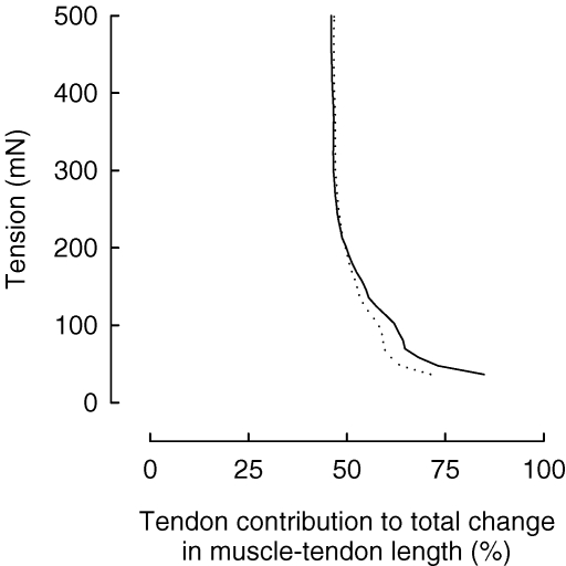
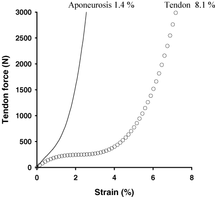
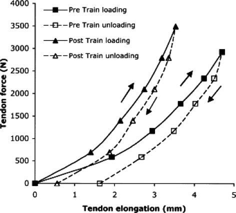
Tendon properties contribute to the complex interaction of the central nervous system, muscle-tendon unit and bony structures to produce joint movement. Until recently limited information on human tendon behaviour in vivo was available; however, novel methodological advancements have enabled new insights to be gained in this area. The present review summarizes the progress made with respect to human tendon and aponeurosis function in vivo, and how tendons adapt to ageing, loading and unloading conditions. During low tensile loading or with passive lengthening not only the muscle is elongated, but also the tendon undergoes significant length changes, which may have implications for reflex responses. During active loading, the length change of the tendon far exceeds that of the aponeurosis, indicating that the aponeurosis may more effectively transfer force onto the tendon, which lengthens and stores elastic energy subsequently released during unloading, in a spring-like manner. In fact, data recently obtained in vivo confirm that, during walking, the human Achilles tendon provides elastic strain energy that can decrease the energy cost of locomotion. Also, new experimental evidence shows that, contrary to earlier beliefs, the metabolic activity in human tendon is remarkably high and this affords the tendon the ability to adapt to changing demands. With ageing and disuse there is a reduction in tendon stiffness, which can be mitigated with resistance exercises. Such adaptations seem advantageous for maintaining movement rapidity, reducing tendon stress and risk of injury, and possibly, for enabling muscles to operate closer to the optimum region of the length-tension relationship.
Figures


References
-
- Alexander RM. Energy-saving mechanisms in walking and running. J Exp Biol. 1991;160:55–69. - PubMed
-
- Alexander RM, Bennet-Clark HC. Storage of elastic strain energy in muscle and other tissues. Nature. 1977;265:114–117. - PubMed
-
- Arampatzis A, Stafilidis S, DeMonte G, Karamanidis K, Morey-Klapsing G, Bruggemann GP. Strain and elongation of the human gastrocnemius tendon and aponeurosis during maximal plantarflexion effort. J Biomech. 2005;38:833–841. - PubMed
-
- Arndt AN, Bruggemann GP, Koebke J, Segesser B. Asymmetrical loading of the human tricpes surae. I. Mediolateral force difference in the Achilles tendon. Foot Ankle Int. 1999;20:445–449. - PubMed
-
- Arnoczky SP, Lavagnino M, Whallon JH, Hoonjan A. In situ cell nucleus deformation in tendons under tensile load; a morphological analysis using confocal laser microscopy. J Orthop Res. 2002;20:29–35. - PubMed
Publication types
MeSH terms
LinkOut - more resources
Full Text Sources
Medical