. 2007 Dec;103(5):1855-63.
doi: 10.1111/j.1471-4159.2007.04918.x. Epub 2007 Sep 13.

Neurexin Ibeta and neuroligin are localized on opposite membranes in mature central synapses

Affiliations

Neurexin Ibeta and neuroligin are localized on opposite membranes in mature central synapses

Otto Berninghausen et al. J Neurochem. 2007 Dec.

Abstract

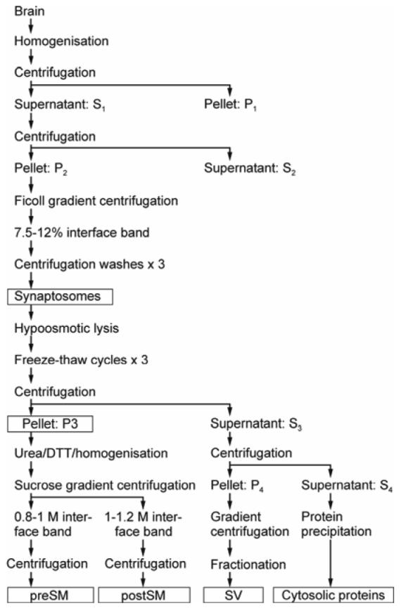
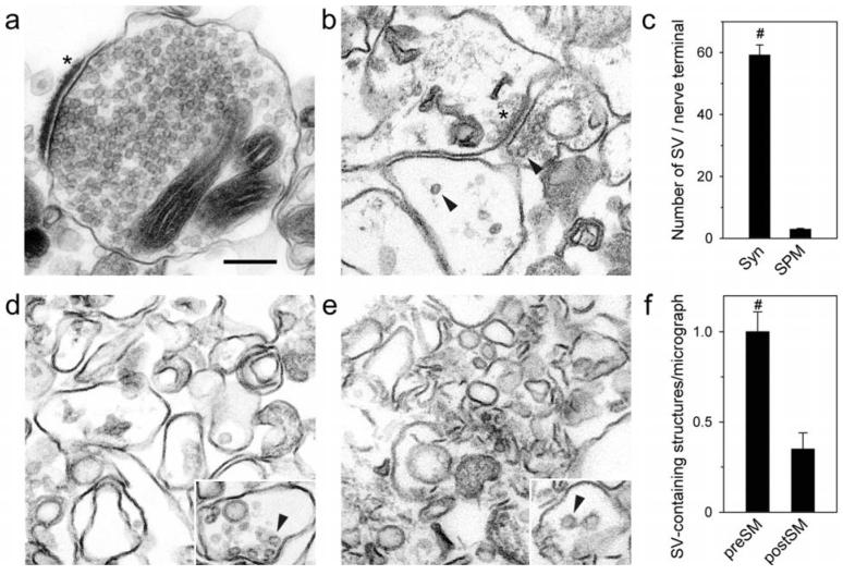
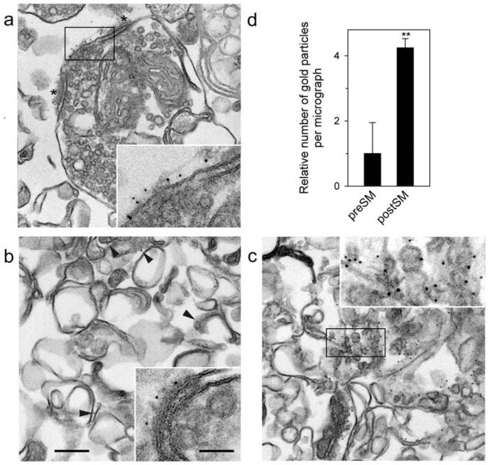
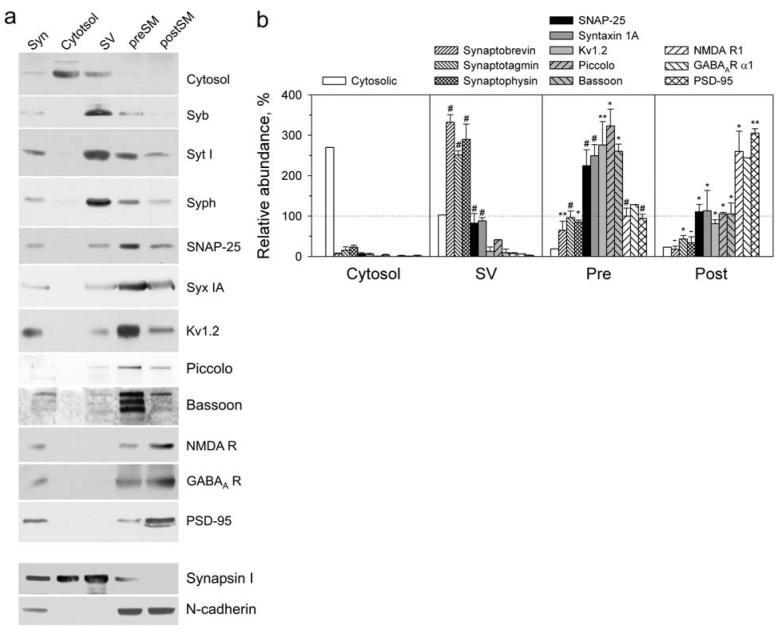
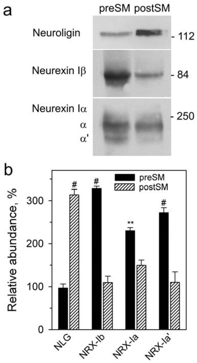
Synaptogenesis requires formation of trans-synaptic complexes between neuronal cell-adhesion receptors. Heterophilic receptor pairs, such as neurexin Ibeta and neuroligin, can mediate distinct intracellular signals and form different cytoplasmic scaffolds in the pre- and post-synaptic neuron, and may be particularly important for synaptogenesis. However, the functions of neurexin and neuroligin depend on their distribution in the synapse. Neuroligin has been experimentally assigned to the post-synaptic membrane, while the localization of neurexin remains unclear. To study the subcellular distribution of neurexin Ibeta and neuroligin in mature cerebrocortical synapses, we have developed a novel method for the physical separation of junctional membranes and their direct analysis by western blotting. Using urea and dithiothreitol, we disrupted trans-synaptic protein links, without dissolving the lipid phase, and fractionated the pre- and post-synaptic membranes. The purity of these fractions was validated by electron microscopy and western blotting using multiple synaptic markers. A quantitative analysis has confirmed that neuroligin is localized strictly in the post-synaptic membrane. We have also demonstrated that neurexin Ibeta is largely (96%) pre-synaptic. Thus, neurexin Ibeta and neuroligin normally form trans-synaptic complexes and can transduce bidirectional signals.


Figures

Fig. 1
Fig. 1
Schematic diagram of separation and fractionation of synaptic membranes. Details are described under Materials and Methods. Subcellular fractions used in this work are boxed.
Fig. 2
Fig. 2
Morphology of preSM and postSM. Samples from purification steps were aldehyde-fixed and post-fixed with osmium, treated with tannic acid, dehydrated and embedded in Epon. Sectioned specimens were stained with lead citrate and analyzed by EM. Morphological analysis of synaptosomes (a) and P3 (b). Asterisks denote postsynaptic membranes with PSD attached, residual SV are marked with arrowheads. (c) Number of SV found in synaptosomes (Syn) and in identifiable nerve terminals in P3 (n = 5, total number of SV 5,222 in 88 structures for synaptosomes and 207 in 70 structures for P3). (d-e) Representative images of preSM (d) and postSM (e). Insets show examples of structures containing remaining SV (arrowheads). (f) Relative number of vesicle-containing structures in preSM (n = 7; total number of structures 64) and postSM (n = 6; total number of structures 19). Student's t-tests compare the two values, and the results are indicated above; #, p < 0.001. Scale bar for all micrographs and insets, 0.2 μm.
Fig. 3
Fig. 3
Immuno-EM of preSM and postSM. Synaptosomes (a), preSM (b) and postSM (c) membranes were incubated with anti-PSD-95 mAb, fixed, labeled with 5-nm immunogold, before post-fixation and embedding, as in Fig. 2. Insets are magnified views of: (a and c) the respective boxed regions; (b) a rare example of postsynaptic membrane remaining in preSM. Note the immunogold label on postsynaptic membranes present in synaptosomes, remaining to some extent in preSM and abundant in postSM (arrowheads). Asterisks mark PSD in (a). Scale bar, 0.2 μm (0.5 μm for all insets). (d) Quantification of PSD-95-immunogold particles in the preSM and postSM fractions, relative to their contents in preSM. The data are the means of several experiments shown together with SEM (n = 7, total number of gold particles found: 245 in preSM and 1,349 in postSM). Student's t-test compares the amount of PSD-95 in each fraction, and the result is indicated above; **, p < 0.01.
Fig. 4
Fig. 4
Enrichment of synaptic proteins in respective fractions. (a) Representative western blots of subcellular brain fractions (syn, synaptosomes), stained for marker proteins. The fractions, containing 50 μg protein, were prepared as described under Supplementary Methods online, separated by SDS-electrophoresis in 6-10% polyacrylamide gels and transferred onto polyvinylidene fluoride membranes. Proteins were immunostained using respective primary and secondary antibodies and visualized by chemiluminescence with digital imaging. The following marker proteins were used: SV (synaptophysin, Syph; synaptotagmin I, Syt; and synaptobrevin, Syb); presynaptic membranes (syntaxin IA, Syx IA; SNAP-25; voltage-dependent potassium channel, Kv1.2; piccolo and bassoon); postsynaptic membranes (NMDA receptor 1, GABAA receptor α1 and PSD-95) and wider distribution (synapsin I and N-cadherin). (b) Quantification of western blots. The graph shows the relative amounts of each protein in the cytosol, preSM and postSM compared to the same protein's content in synaptosomes (taken to be 100 %). The data are the means of several experiments (n = 3-5) together with SEM. Student's t-tests compare the amount of each protein in a fraction relative to that in the adjacent fraction on the left, and the results are indicated above each value; *, p < 0.05; **, p < 0.01; #, p < 0.001; -, non-significant.
Fig. 5
Fig. 5
Analysis of the trans-synaptic distribution of neuroligin, neurexin Iβ and neurexin Iα. (a) A representative western blot of the preSM and postSM fractions stained with anti-neuroligin and anti-neurexin antibodies. Letters on the left denote the two isoforms of neurexin Iα. Positions and molecular masses (kDa) of marker proteins are shown on the right. (b) Quantification of western blots as in (a) (n = 3). The graph shows the relative amounts (±SEM) of each protein in the preSM and postSM, expressed as a per cent of its content in synaptosomes. NLG, neuroligin; NRX, neurexin. The results of Student's t-test compare the amounts of each protein in the two fractions and are shown near the values; #, p < 0.001; **, p < 0.01.

Similar articles

Cited by

References

    1. Boucard AA, Chubykin AA, Comoletti D, Taylor P, Sudhof TC. A splice code for trans-synaptic cell adhesion mediated by binding of neuroligin 1 to alpha- and beta-neurexins. Neuron. 2005;48:229–236. - PubMed
    1. Chih B, Gollan L, Scheiffele P. Alternative splicing controls selective trans-synaptic interactions of the neuroligin-neurexin complex. Neuron. 2006;51:171–178. - PubMed
    1. Chubykin AA, Liu X, Comoletti D, Tsigelny I, Taylor P, Sudhof TC. Dissection of synapse induction by neuroligins: effect of a neuroligin mutation associated with autism. J. Biol. Chem. 2005;280:22365–22374. - PubMed
    1. Chung YH, Shin C, Kim MJ, Lee BK, Cha CI. Immunohistochemical study on the distribution of six members of the Kv1 channel subunits in the rat cerebellum. Brain Res. 2001;895:173–177. - PubMed
    1. Cohen RS, Blomberg F, Berzins K, Siekevitz P. The structure of postsynaptic densities isolated from dog cerebral cortex. I. Overall morphology and protein composition. J. Cell Biol. 1977;74:181–203. - PMC - PubMed

Publication types

MeSH terms