Comparative Study
. 2007 Sep 14:7:10.
doi: 10.1186/1472-6793-7-10.

Inflammatory cytokines induce MAdCAM-1 in murine hepatic endothelial cells and mediate alpha-4 beta-7 integrin dependent lymphocyte endothelial adhesion in vitro

Affiliations
Comparative Study

Inflammatory cytokines induce MAdCAM-1 in murine hepatic endothelial cells and mediate alpha-4 beta-7 integrin dependent lymphocyte endothelial adhesion in vitro

Tomoaki Ando et al. BMC Physiol. .

Abstract

Background: MAdCAM-1 plays a central role in T-lymphocyte homing to the gut, but its role in chronic liver inflammation remains unknown. Therefore, this study measured MAdCAM-1 expression, regulation, and function in cultured murine hepatic endothelial cells.

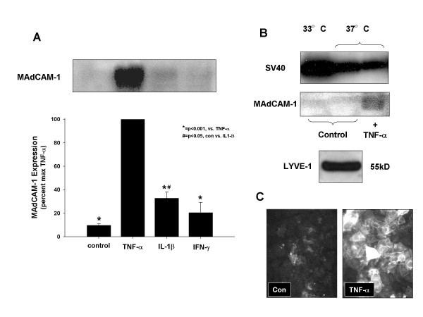
Methods: Cultures of hepatic endothelial cells (HEC) were prepared from mice expressing a temperature-sensitive SV40 large T antigen (H-2Kb-tsA58) under the control of an IFN-gamma promoter. Time and dose dependent expression of MAdCAM-1 in response to TNF-alpha, IL-1 beta and IFN-gamma was studied by immunoblotting. Lymphocyte adhesion was studied using alpha 4 beta 7 integrin expressing lymphocytes (TK-1) +/- anti-MAdCAM-1 mAb.

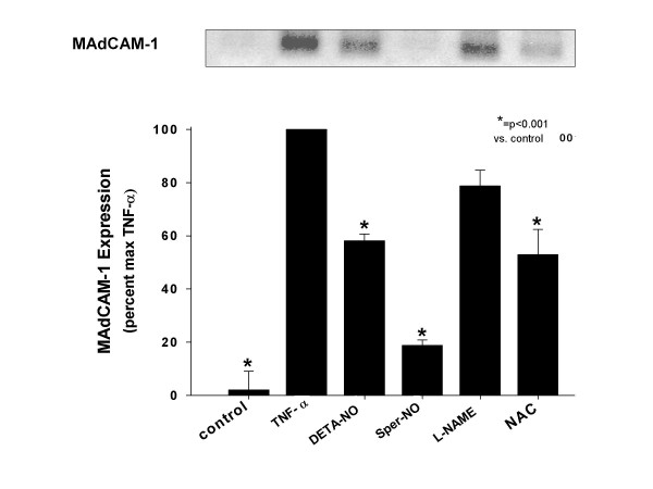
Results: TNF-alpha induced MAdCAM-1 dose-and time-dependently with maximum expression at 20 ng/ml and at 48 hours. IL-1 beta also induced MAdCAM-1 to a lesser extent compared to TNF-alpha; IFN-gamma did not induce MAdCAM-1. TNF-alpha significantly increased lymphocyte-endothelial adhesion (P < 0.01), which was reversed by anti-MAdCAM-1 antibody. MAdCAM-1 expression was also reduced by N-acetylcysteine and by two NO donors (SperNO, DETANO) suggesting that hepatic endothelial MAdCAM-1 is oxidant and NO regulated.

Conclusion: MAdCAM-1 is a major determinant of leukocyte recruitment in chronic inflammation and is expressed by HEC in response to IL-1 beta and TNF-alpha. This system may provide a useful model for studying inflammatory mechanisms in liver disease and help determine if controlled MAdCAM-1 expression might influence inflammation in liver disease.


Figures

Figure 1
Figure 1
Establishment of hepatic endothelial cells (HEC). A. Phase contrast image of HEC. B. Immunofluorescent staining for VCAM-1 in HEC. C. Immunofluoresent images of mouse endothelial cell antigen-32 (MECA-32) expressed on the surface of HEC monolayers. D. Distribution of DiI-labeled acetylated low density lipoprotein -(LDL), an endothelial cell specific biomarker incorporated into endothelial cells by receptor mediated endocytosis.
Figure 2
Figure 2
MAdCAM-1 is expressed by cytokine activated hepatic endothelial cells. A. Hepatic endothelial cells were treated with TNF-α (20 ng/ml), or IL-1β (10 ng/ml) or IFN-γ (1000 u/ml) for 24 h. B. Immunoblotting for SV40 Large-T antigen and MAdCAM-1 C. Immunostaining for MAdCAM-1 on the HEC cell surface.
Figure 3
Figure 3
TNF-α stimulates MAdCAM-1 expression in a dose-and time- dependent manner. A. Confluent HEC endothelial cells were incubated for 24 h with increasing concentrations of mouse TNF-α (as indicated), and MAdCAM-1 induction (measured as western blotting density), expressed as a percent of the maximum, (set as the scan density in 24 h, 20 ng/ml TNF-α treated HEC, [100%]). MAdCAM-1 Expression is TNF-α concentration dependent B. HEC were incubated with 20 ng/mL TNF-α for time periods up to 48 h (as indicated). MAdCAM-1 protein expression was time dependently increased at 12, 24 and 48 h. (Max expression [100%] in these experiments was determined as the blotting scan density seen at 48 hours). Expression at T = 0 represents MAdCAM-1 expressed by un-stimulated hepatic endothelial cells. Each value represents the mean ± s.e.; each group (n = 3). *P < 0.01, **P < 0.01 vs. TNF-α treatment.
Figure 4
Figure 4
Quantitation of cells expressing MAdCAM-1 protein measured by flow cytometry A. MAdCAM-1 expression 24 h after TNF-α stimulation (20 ng/ml) B. Time course analysis of MAdCAM-1 expression. The concentration of TNF-α in this study was 20 ng/ml. Values represent means ± SE; n = 3 experiments in each group.
Figure 5
Figure 5
Effect of nitric oxide (NO) donors and N-acetylcysteine, an antioxidant on MAdCAM-1 expression by HEC in response to TNF-α. HEC were pretreated for 30 minutes with the NO donors, DETA-NO or SperNO (500 uM), or L-NAME (1 mM), or N-acetylcysteine (1 mM). Tumor necrosis factor (TNF)-α (20 ng/ml) was then added to these cultures for 48 h. MAdCAM-1 expression was significantly increased by TNF-α and significantly reduced by DETA-NO, SperNO or NAC, but not by L-NAME. Values represent the mean ± SE; n = 3 experiments in each group. * = p < 0.01 vs. TNF-α treatment.
Figure 6
Figure 6
TNF-α induces MAdCAM-1 dependent adhesion of TK-1 lymphocytes to HEC. Compared to controls which exhibited 57.68 ± 3.77% of the maximum lymphocyte adhesion, 20 ng/ml TNF-α (24 h), induced a significant increase in TK-1 cell adhesion (set as maximal '100%' adhesion). The increased adhesion induced by TNF-α was significantly reduced to 84.8 ± 3.99% of maximal adhesion by pre-/co-treatment of HEC with the blocking mouse MAdCAM-1 Ab 'MECA-367'(10 ug/ml). The binding but not blocking MAdCAM-1 antibody MECA-89 did not significantly reduce TNF-α induced adhesion (98 ± 5.25% maximal adhesion). Each value represents the mean ± s.d.; each group (n = 6).

Similar articles

Cited by

References

    1. Picker LJ, Butcher EC. Physiological and molecular mechanisms of lymphocyte homing. Annu Rev Immunol. 1992;10:561–591. doi: 10.1146/annurev.iy.10.040192.003021. - DOI - PubMed
    1. Connor EM, Eppihimer MJ, Morise Z, Granger DN, Grisham MB. Expression of mucosal addressin cell adhesion molecule-1 (MAdCAM-1) in acute and chronic inflammation. J Leukoc Biol. 1999;65:349–355. - PubMed
    1. Picarella D, Hurlbut P, Rottman J, Shi X, Butcher E, Ringler DJ. Monoclonal antibodies specific for beta 7 integrin and mucosal addressin cell adhesion molecule-1 (MAdCAM-1) reduce inflammation in the colon of scid mice reconstituted with CD45RBhigh CD4+ T cells. J Immunol. 1997;158:2099–2106. - PubMed
    1. Jaeschke H. Cellular adhesion molecules: regulation and functional significance in the pathogenesis of liver diseases. Am J Physiol. 1997;273:G602–G611. - PubMed
    1. Steinhoff G, Behrend M, Schrader B, Pichlmayr R. Intercellular immune adhesion molecules in human liver transplants: overview on expression patterns of leukocyte receptor and ligand molecules. Hepatology. 1993;18:440–453. - PubMed

Publication types

LinkOut - more resources