. 2007 Sep 14:8:76.
doi: 10.1186/1471-2199-8-76.

HAP1 can sequester a subset of TBP in cytoplasmic inclusions via specific interaction with the conserved TBP(CORE)

Affiliations

HAP1 can sequester a subset of TBP in cytoplasmic inclusions via specific interaction with the conserved TBP(CORE)

Justin R Prigge et al. BMC Mol Biol. .

Abstract

Background: Huntington's disease, spinal and bulbar muscular atrophy, and spinocerebellar ataxia 17 (SCA17) are caused by expansions in the polyglutamine (polyQ) repeats in Huntingtin protein (Htt), androgen receptor protein (AR), and TATA-binding protein (TBP), respectively. Htt-associated protein 1 (HAP1), a component of neuronal cytoplasmic stigmoid bodies (STBs), can sequester polyQ-expanded Htt and AR in STBs, thereby antagonizing formation of the nuclear aggregates associated with apoptotic neuron loss and disease progression.

Results: Clones of HAP1 were isolated from unbiased two-hybrid screens for proteins that interact with TBP. Domain mapping showed that regions between amino acids 157 and 261 and between amino acids 473 and 582 of mouse HAP1 both bind specifically to the conserved C-terminal TBP(CORE) domain, away from the TBP N-terminal polyQ region. When fluorescently tagged versions of HAP1 or TBP were expressed independently in COS-7, 293, or Neuro-2a cells, all TBP localized to the nucleus and all HAP1 assembled into cytoplasmic stigmoid-like bodies (STLBs). When co-expressed, a portion of the TBP was assembled into the HAP1 STLBs while the remainder was localized to the nucleus. Although the TBP N terminus, including the polyQ region, was unnecessary for TBP-HAP1 interaction, in mammalian cells, removal of the TBP Q(repeat) reduced the proportion of TBP that assembled into STLBs, whereas expansion of the Q(repeat) had no significant affect on TBP subcellular localization.

Conclusion: HAP1 can sequester a subset of TBP protein away from the nucleus; extranuclear TBP sequestration is quantitatively influenced by the TBP polyQ repeat. These results suggest HAP1 could provide protection from SCA17 neuropathology.


Figures

Figure 1
Figure 1
Yeast two-hybrid bait constructs, prey libraries, and screens. (A) Bait constructs. TBP-FL, TBP-C, and TBP-N were expressed from the pDBLeu plasmid, which fused the Gal4 DNA-binding domain (DBD) upstream of TBP. N and C designate the vertebrate-specific N terminus and the pan-eukaryotic TBPCORE, respectively. (B) Three prey libraries were constructed and inserted into pPC86, which fused prey cDNAs downstream of the Gal4 activation domain (AD). Characteristics of each library are indicated. Libraries were constructed from oligo(dt)-primed placental and pregnant uteri RNA or from random-primed placenta + pregnant uteri RNA (p+u). Below is shown PCR analysis of arbitrary clones from both of the oligo(dT)-primed libraries using a primer pair that spans the multiple cloning site of the vector. Lane "λ" contained Hind III/Eco RI-cut λ-phage DNA markers. Landmark band sizes are indicated at the left of each gel; the asterisk denotes the size of the PCR product arising from empty prey vector. (C) The results of the two yeast two-hybrid screens performed are shown. In both screens, TBP-FL was used as bait to screen either the oligo(dT)- or random-primed placenta + uteri prey libraries for interacting proteins. TBP-interacting prey library clones were subsequently identified by sequencing.
Figure 2
Figure 2
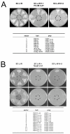
Protein interactions in the yeast two-hybrid system. Bait and prey plasmid combinations within individual yeast clones are indicated below. Sector designations correspond to those on plates. Growth of yeast on SC-L-W indicated that clones contained both the bait and prey plasmid. (A) Interaction with TBP-FL, TBP-N, and TBP-C. Sectors 1–3 show the interaction of HAP1-B155–582 with TBP-FL, TBP-C, or TBP-N, respectively. The interaction of HAP1 with TBP-FL and TBP-C, but not TBP-N resulted in growth under all conditions tested. Sectors 4–6 were auto-activation controls for TBP baits. Sectors 7–8 contained strong and weak positive interaction controls, respectively, that were supplied with the ProQuest system. The strong interaction control grew well under all conditions tested. The weak interaction control did not grow on SC-L-W-H-U, suggesting that this condition allowed growth of yeast containing only strong bait-prey interactions. Sector 9 was a negative auto-activation control showing that yeast bearing only the empty bait vector and an empty prey plasmid (no inserts) did not grow under conditions that selected for interacting bait-prey protein pairs. (B) Finer TBP deletions. Sectors 1–6 show the interaction of HAP1-A7–598 with TBP-ΔQ (sector 1) and truncated TBP-C clones (as indicated in table below; sectors 2–6). HAP1 interacted with TBP-ΔQ under all conditions tested (sector 1), but did not interact with any of the truncated TBP-C clones (sectors 2–6), suggesting that an intact TBPCORE is required for the HAP1-TBP interaction to occur. Sectors 7–12 were auto-activation controls for TBP baits, of which none were auto-active.
Figure 3
Figure 3
Co-immunoprecipitation assay. TBP/HAP1-B155–582 interactions in co-transfected cells. (A) 293 cells were co-transfected with pCMV-HA-TBP-FL and pCMV-MYC-HAP1. Whole cell lysates (WCL) (lanes 1–2, where lane 1 and lane 2 represent 4% of each WCL used for immunoprecipitation in lanes 3 and 4, respectively) or immunoprecipitated samples (lanes 3–4) were assayed using western blots with anti-MYC antibody. MYC-tagged HAP1 co-precipitated with TBP in the presence of the anti-TBP antibody (lane 3), but not with the non-specific antibody (lane 4). (B) COS-7 cells were co-transfected with pFLAG-CMV-GFP-TBP-FL (lanes 1–4, 7–8) or pFLAG-CMV-GFP (lanes 5–6, 9) and pCMV-MYC-HAP1-B155–582 (lanes 1–9). Whole cell lysates (lanes 1, 3, and 5, HAP1) or immunoprecipitated samples (lanes 2, 4, and 6, HAP1) were assayed using western blots and anti-MYC antibody. MYC-tagged HAP1 co-precipitated with FLAG-tagged-GFP-TBP in the presence of the anti-FLAG antibody (lane 2), but not with the non-specific antibody (lane 4). Myc-tagged HAP1 did not co-precipitate with FLAG-tagged-GFP in the presence of anti-FLAG antibody (lane 6). 30% of each immunoprecipitated sample was also blotted with anti-FLAG antibody (lanes 7–9) to verify the amount of FLAG-tagged protein that was captured in each co-precipitation assay (lanes 7–9 represent co-precipitated samples in lanes 2, 4, and 6, respectively).
Figure 4
Figure 4
Identification of TBP/HAP1 interaction domains. The interaction of truncated HAP1 clones with TBP-C, TBP-N, and auto-activation controls are shown. Yeast containing the indicated bait and prey plasmids (listed below) were grown on SC-L-W (top panel), which selects for the presence of the bait and prey plasmids, and SC-L-W-H + 50 mM 3-AT (bottom panel), which selects for the interaction between bait and prey. All baits were expressed from the pDBLeu plasmid and all truncated HAP1 preys were expressed from the pPC86 plasmid. Sectors 3, 6, 9, 12, 15, 18, 21, 24, 27, 30, 33, 36, and 39 were controls confirming that truncated HAP1 clones were not auto-active. Sectors 2, 5, 8, 11, 14, 17, 20, 23, 26, 29, 32, 35, and 38 confirmed that none of the HAP1 clones interacted with TBP-N. Clones 10, 13, 19, and 22 did not grow on SC-L-W-H + 50 mM 3-AT, which suggested that these clones lacked the TBP interaction domain, whereas growth of clones 1, 4, 7, 16, 25, 28, 31, 34, and 37 on SC-L-W-H + 50 mM 3-AT suggested that these clones contained a TBP interaction domain. Three independent clones of all HAP1 truncations were tested and each gave the same result; one representative set is shown.
Figure 5
Figure 5
Deletion analysis of the TBP interacting regions on HAP1. (A) The full-length HAP1-B protein is represented at top, including the location of the known acidic domain (AcD) and the two coiled coil domains. HAP1 amino acids encoded by each cDNA clone in pPC86 are shown at left. The presence (+) or absence (-) of interaction with TBP-C is shown at right. Results suggested that the TBP interacting regions on HAP1 were between amino acids 157 and 261, and between amino acids 473 and 582. (B) Amino acid sequences of HAP1 TBP-interacting regions.
Figure 6
Figure 6
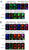
TBP and HAP1 co-localization assay in COS-7, 293, and Neuro-2a cells. (A) cells transfected with a single expression plasmid, as indicated at top. Forty-eight hours after transfection, cells were stained with Hoechst 33342 and fluorescent photomicrographs of the blue, red, and green channels were taken. Hoechst stain (blue) shows nuclei. CMVp-GFP-HAP1-A7–598 was exclusively cytoplasmic in all cells tested, where it assembled into strongly fluorescent STLBs. CMVp-DSRed2-TBP was exclusively nuclear with heterogenous subnuclear distributions when expressed alone. The negative controls, CMVp-DSRed2-PTIP, MED15, and U2AF65 (encoding nuclear proteins unrelated to TBP, see text), were also localized entirely to the nucleus when expressed alone. (B) co-expression of HAP1 with TBP, PTIP, MED15, or U2AF65. Co-expression of CMVp-GFP-HAP1 and CMVp-DSRed2-TBP shows that all HAP1 remained cytoplasmic, where it assembled into STLBs; however, TBP localization was altered. Thus, whereas much of the TBP still localized to the nucleus, a portion was sequestered into GFP-HAP1-STLBs. White arrows designate representative examples of extranuclear TBP in STLBs. As negative controls, DSRed2-PTIP, MED15, and U2AF65 were not detected in GFPHAP1-STLBs.
Figure 7
Figure 7
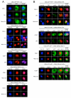
HAP1 and TBP-FL, -ΔQ, or -Q50 co-localization assays in COS-7 and Neuro-2a cells. (A) Top two rows show cells transfected with a single expression plasmid: CMVp-GFP-TBP-FL (at left) and CMVp-DSRed2-HAP1-B155–582 (at right). GFP-TBP was exclusively nuclear and RFP-HAP1 assembled into cytoplasmic STLBs. Bottom two rows show co-transfected cells containing GFP-TBP and RFP-HAP1. Upon co-expression with HAP1, a portion of GFP-TBP became localized to the cytoplasmic, RFP-HAP1-STLBs. (B) Top two rows show cells transfected with a single expression plasmid: CMVp-DSRed2-TBP-ΔQ (at left) and CMVp-DSRed2-TBP-Q50 (at right). Both RFP-TBP-ΔQ and RFP-TBP-Q50 proteins localized exclusively to the nucleus when expressed alone. Cells co-transfected with GFP-HAP1-A7–598 and either RFP-TBP-ΔQ (middle two rows) or RFP-TBP-Q50 (bottom two rows) showed altered localization of TBP away from the nucleus. RFP-TBP-ΔQ, which lacks the polyQ repeat, and RFP-TBP-Q50, which contains an expanded polyQ repeat, each were partially localized in the GFP-HAP1-STLBs.
Figure 8
Figure 8
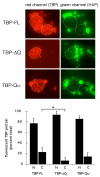
PolyQ dependence of relative nuclear and cytoplasmic TBP protein levels. (A) representative fluorescent photomicrographs used for quantitative analyses. Cells were transfected with CMVp-GFP-HAP1-A7–598 and either CMVp-DSRed2-TBP-FL, CMVp-DSRed2-TBP-ΔQ, or CMVp-DSRed2-TBP-Q50, as indicated at left. White dotted line delineates nuclei in each frame. (B) nuclear-cytoplasmic TBP distributions. Bars show average plus one standard deviation. Aserisks, cytoplasmic levels of TBP-ΔQ were significantly less than those of TBP-FL, P < 0.05. Cytoplasmic levels of TBP-FL and TBP-Q50 were not significantly different.

Similar articles

Cited by

References

    1. Everett CM, Wood NW. Trinucleotide repeats and neurodegenerative disease. Brain. 2004;127:2385–2405. doi: 10.1093/brain/awh278. - DOI - PubMed
    1. MacDonald ME, Ambrose CM, Duyao MP, Myers RH, Lin C, Srinidhi L, Barnes G, Taylor SA, James M, Groot N, MacFarlane H, Jenkins B, Anderson MA, Wexler NS, Gusella JF, Bates GP, Baxendale S, Hummerich H, Kirby S, North M, Youngman S, Mott R, Zehetner G, Sedlacek Z, Poustka A, Frischauf A-M, Lehrach H, Buckler AJ, Church D, Doucette-Stamm L, et al. A novel gene containing a trinucleotide repeat that is expanded and unstable on Huntington's disease chromosomes. The Huntington's Disease Collaborative Research Group. Cell. 1993;72:971–983. doi: 10.1016/0092-8674(93)90585-E. - DOI - PubMed
    1. Landles C, Bates GP. Huntingtin and the molecular pathogenesis of Huntington's disease. Fourth in molecular medicine review series. EMBO Rep. 2004;5:958–963. doi: 10.1038/sj.embor.7400250. - DOI - PMC - PubMed
    1. Li SH, Hosseini SH, Gutekunst CA, Hersch SM, Ferrante RJ, Li XJ. A human HAP1 homologue. Cloning, expression, and interaction with huntingtin. J Biol Chem. 1998;273:19220–19227. doi: 10.1074/jbc.273.30.19220. - DOI - PubMed
    1. Li XJ, Li SH, Sharp AH, Nucifora FC, Jr, Schilling G, Lanahan A, Worley P, Snyder SH, Ross CA. A huntingtin-associated protein enriched in brain with implications for pathology. Nature. 1995;378:398–402. doi: 10.1038/378398a0. - DOI - PubMed

Publication types

MeSH terms