. 2007 Sep 14:4:36.
doi: 10.1186/1742-4682-4-36.

Cable properties and propagation velocity in a long single chain of simulated myocardial cells

Affiliations

Cable properties and propagation velocity in a long single chain of simulated myocardial cells

Lakshminarayanan Ramasamy et al. Theor Biol Med Model. .

Abstract

Background: Propagation of simulated action potentials (APs) was previously studied in short single chains and in two-dimensional sheets of myocardial cells 123. The present study was undertaken to examine propagation in a long single chain of cells of various lengths, and with varying numbers of gap-junction (g-j) channels, and to compare propagation velocity with the cable properties such as the length constant (lambda).

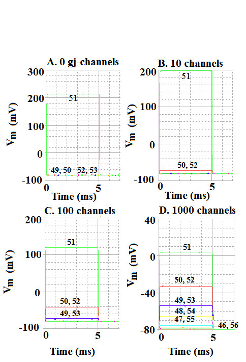
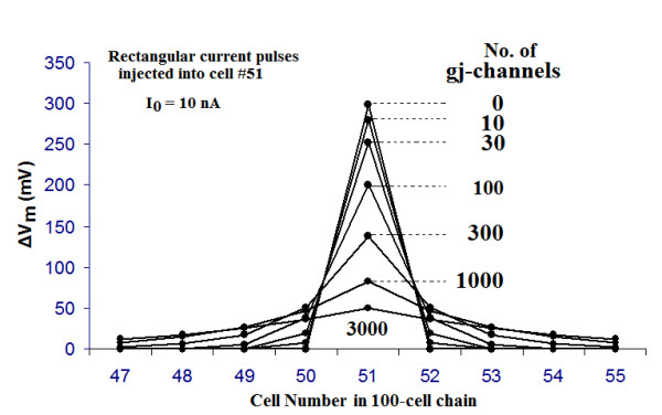
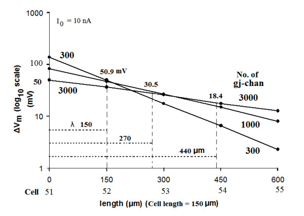
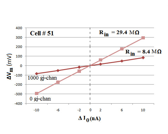
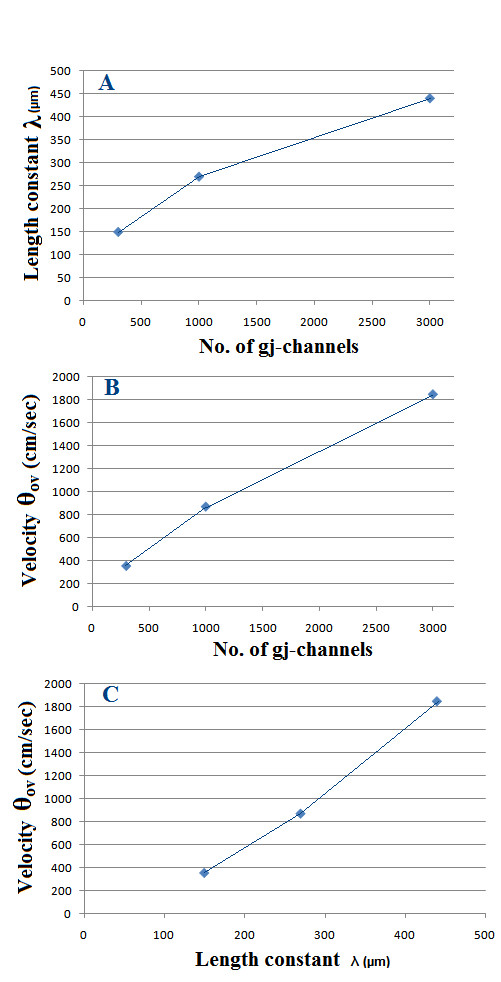
Methods and results: Simulations were carried out using the PSpice program as previously described. When the electric field (EF) mechanism was dominant (0, 1, and 10 gj-channels), the longer the chain length, the faster the overall velocity (theta(ov)). There seems to be no simple explanation for this phenomenon. In contrast, when the local-circuit current mechanism was dominant (100 gj-channels or more), theta(ov) was slightly slowed with lengthening of the chain. Increasing the number of gj-channels produced an increase in theta(ov) and caused the firing order to become more uniform. The end-effect was more pronounced at longer chain lengths and at greater number of gj-channels. When there were no or only few gj-channels (namely, 0, 10, or 30), the voltage change (DeltaV(m)) in the two contiguous cells (#50 & #52) to the cell injected with current (#51) was nearly zero, i.e., there was a sharp discontinuity in voltage between the adjacent cells. When there were many gj-channels (e.g., 300, 1000, 3000), there was an exponential decay of voltage on either side of the injected cell, with the length constant (lambda) increasing at higher numbers of gj-channels. The effect of increasing the number of gj-channels on increasing lambda was relatively small compared to the larger effect on theta(ov). theta(ov) became very non-physiological at 300 gj-channels or higher.

Conclusion: Thus, when there were only 0, 1, or 10 gj-channels, theta(ov) increased with increase in chain length, whereas at 100 gj-channels or higher, theta(ov) did not increase with chain length. When there were only 0, 10, or 30 gj-channels, there was a very sharp decrease in DeltaV(m) in the two contiguous cells on either side of the injected cell, whereas at 300, 1000, or 3000 gj-channels, the voltage decay was exponential along the length of the chain. The effect of increasing the number of gj-channels on spread of current was relatively small compared to the large effect on theta(ov).


Figures

Figure 1
Figure 1
Schematic diagram for the single chain of 100 myocardial cells. Rows 2 (B), 3 (C), 4 (D), 8(H), and 9 (I) have been omitted in order to contain the size of the figure. For the propagation velocity experiment, cell #1 was stimulated intracellularly with rectangular depolarizing current pulses of 0.1 nA amplitude and 0.25 ms duration. The resultant action potentials (APs) were recorded only from cells 1, 10, 20, 30, 40, 50, 60, 70, 80, 90, and 100 in order to limit the number of traces. For the length constant experiments, intracellular depolarizing rectangular current pulses (10 nA, 5 ms) were applied to cell #51 (middle of chain), and the resulting voltage changes in cell #51 and its immediate neighbors were measured. For this type of experiment, the cells were made inexcitable by removing their GTABLEs.
Figure 2
Figure 2
Propagation of simulated action potentials (APs) in a single linear chain of 100 myocardial cells. Cell #1 was stimulated intracellularly, and the resultant APs were recorded from only cells #1, 10, 20, 30, 40, 50, 60, 70, 80, 90, and 100 (to limit the number of traces). The number of gap-junction (g-j) channels at the cell junctions was varied over a wide range, but only five are illustrated, namely 0 gj-channels (A), 300 (B), 1,000 (C), 3,000 (D), and 10,000. The traces numbered in panel A are for APs recorded from cells #1, 10, 20, and 100; the remaining traces are bunched up between cells 20 and 100, some of them being nearly superimposed. Note that adding gj-channels markedly speeds up the velocity of propagation. In panel E, all 11 traces are superimposed.
Figure 3
Figure 3
Graphic summary that quantitates how the propagation velocity in simulated cardiac APs varies with the number of gj-channels in the single chain of 100 cells. Note that relationship is nearly linear up to 300 gj-channels. Increasing the number of gj-channels 10-fold (from 100 to 1,000) increased velocity about 5-fold (5.2 fold).
Figure 4
Figure 4
Graphic summary that quantitates how the overall propagation velocity varies with the length of the single chain. For this experiment, the 100-cell chain was shortened to 50, 20, and 10 cells, so that the results could be reliably compared, and the number of gj-channels in each chain length was varied from 0 to 100. The results show that when there was no or only few gj-channels (namely, 0, 1 or 10), propagation velocity increased with length of the chain. In contrast, when there were many gj-channels (e.g., 30, 50, 70, or 100), the velocity was slowed when chain length was increased, the most prominent effect occurring between chain lengths of 10 and 20 cells. As can be seen, the family of curves tended to converge at the chain length of 100 cells.
Figure 5
Figure 5
Experiment to measure the spread of current in the linear chain of 100 cells. The myocardial cells were rendered inexcitable by removing their GTABLEs. Depolarizing current pulses (10 nA, 5 ms) were applied intracellularly to cell #51 near the mid-point of the chain, and the resulting membrane voltage changes were recorded from cell #51 and its immediate neighbors (e.g., cells 44–58). The number of gj-channels was varied over a wide range (namely 0, 10, 30, 100, 300, 1000, and 3000), but the results from only 4 are illustrated in this figure: 0 gj-channels (A), 10 (B), 100 (C), and 1,000 (D). A: With no gj-channels, the voltage change in cell #51 was very large (ca 215 mV), whereas there was almost zero voltage change in the contiguous cells (49, 50, 52, 53). B: with 10 gj-channels, the ΔVm in cell 51 was ca 200 mV, whereas that in cells on either side (cells 50 & 52) was only about 8 mV. C: With 100 gj-channels, the ΔVm in cell 51 was ca 120 mV, that in cells 50 and 52 was ca 40 mV, and that in cells 49 and 53 was ca 8 mV. D: With 1000 gj-channels, the ΔVm in cell 51 was ca 84 mV and those in the contiguous cells were ca 44 mV (cells 50 & 52), ca 26 mV (cells 49 & 53), ca 15 mV (cells 48 & 54), and ca 8 mV (cells 47 & 55).
Figure 6
Figure 6
Graphic summary of the data collected from the experiments on the spread of current with various numbers of gj-channels (0, 10, 30, 100, 300, 1,000, and 3,000). Rectangular current pulses (10 nA, 5 ms) were injected intracellularly into cell #51 (near the middle of the linear chain of 100 cells), and the resulting membrane potential changes (ΔVm) were measured in the injected cell and its immediate neighbors. The myocardial cells were made inexcitable by removal of their GTABLEs. As can be seen, when there were no gj-channels, the ΔVm in the two contiguous cells (50 & 52) was nearly zero. When there were 10 or 30 channels, there was a small ΔVm in cells 50 and 52. When there were 300, 1000, or 3000 channels, the fall-off of ΔVm was exponential, i.e., the cells behaved like a long cable.
Figure 7
Figure 7
The length constant data obtained for 300, 1000, and 3000 gj-channels are plotted on a semi-logarithmic plot to illustrate that the data points form a straight line. The ordinate gives the ΔVm on a log scale, and the abscissa gives the distance along the cable (one direction only) from the point of current injection (middle of cell 51) and assuming the length of each myocardial cell to be 150 μm. Thus, the second labels on the abscissa give the cell number. The value of the length constant (λ) is the distance at which the voltage falls to 1/e (1/2.717) or 36.8 %. Thus, the following λ values were obtained: 150 μm (for 300 gj-channels), 270 μm (for 1000 gj-channels), and 440 μm (for 3000 gj-channels). Hence, increasing the number of gj-channels 10-fold (300 to 3000) increased λ about 3-fold (150 μm to 440 μm).
Figure 8
Figure 8
Voltage/current curves obtained for myocardial cell # 51 near the middle of the single linear 100-cell chain. Depolarizing and hyperpolarizing rectangular current pulses (duration of 5 ms and intensities of 2, 6, and 10 nA) were injected intracellularly into cell #51 and the resultant voltage changes in that cell were recorded and plotted. The ΔV0/I0 curves were linear, in both the depolarizing and hyperpolarizing sectors, because rectification was not incorporated into the basic membrane units that composed each cell. The number of gj-channels connecting the contiguous cells was varied from zero to 3000, but only two of the curves are illustrated, namely for zero and 1,000 gj-channels. As predicted, the curve for 0 gj-channels had a steeper slope and higher input resistance (Rin) than the curve for 1000 gj-channels, namely 29.4 MΩ versus 8.4 MΩ.
Figure 9
Figure 9
Graphic plots for the case where there were many gj-channels (namely 300, 1000, and 3000), giving an exponential fall-off in voltage. A: Length constant (λ) as a function of the number of gj-channels. λ varies approximately with the square root of the number of gj-channels. B: Overall propagation velocity (θov) as a function of the number of channels. C: Velocity (θov) plotted against λ, showing that approximate doubling or tripling of λ produces a greater effect of propagation velocity.

Similar articles

Cited by

  • Adaptive multiscale model for simulating cardiac conduction.
    Hand PE, Griffith BE. Hand PE, et al. Proc Natl Acad Sci U S A. 2010 Aug 17;107(33):14603-8. doi: 10.1073/pnas.1008443107. Epub 2010 Jul 29. Proc Natl Acad Sci U S A. 2010. PMID: 20671202 Free PMC article.
  • Electrophysiology.
    Griffith BE, Peskin CS. Griffith BE, et al. Commun Pure Appl Math. 2013 Dec;66(12):1837-1913. doi: 10.1002/cpa.21484. Epub 2013 Oct 9. Commun Pure Appl Math. 2013. PMID: 36237603 Free PMC article.

References

    1. Sperelakis N, Ramasamy L. Propagation in cardiac muscle and smooth muscle based on electric field transmission at cell junctions: An analysis by PSpice. IEEE-EMB. 2002;21:130. - PubMed
    1. Sperelakis N. Propagation of action potentials between parallel chains of cardiac muscle cells in PSpice simulation. Can J Physiol Pharmacol. 2003;81:1. doi: 10.1139/y03-019. - DOI - PubMed
    1. Sperelakis N, Kalloor B. Transverse propagation of action potentials between parallel chains of cardiac muscle and smooth muscle cells in PSpice simulations. Biomed Eng Online. 2004;3:5. doi: 10.1186/1475-925X-3-5. - DOI - PMC - PubMed
    1. Sperelakis N, Mann J. Evaluation of electric fieldchanges in the cleft between excitable cells. J Theor Biol. 1977;64:71. doi: 10.1016/0022-5193(77)90114-X. - DOI - PubMed
    1. Ramasamy L, Sperelakis N. Effect of transverse gap-junction channels on transverse propagation in an enlarged PSpice model of cardiac muscle. Theor Biol Med Modeling. 2006;3:14. doi: 10.1186/1742-4682-3-14. - DOI - PMC - PubMed

LinkOut - more resources