

Optic disk and nerve fiber layer imaging to detect glaucoma
- PMID: 17868631
- PMCID: PMC2098694
- DOI: 10.1016/j.ajo.2007.07.010
Optic disk and nerve fiber layer imaging to detect glaucoma
Abstract
Purpose: To compare optic disk and retinal nerve fiber layer (RNFL) imaging methods to discriminate eyes with early glaucoma from normal eyes.
Design: Retrospective, cross-sectional study.
Methods: In a tertiary care academic glaucoma center, 92 eyes of 92 subjects (46 with early perimetric open-angle glaucoma and 46 controls) were studied. Diagnostic performance of optical coherence tomography (StratusOCT; Carl Zeiss Meditec, Dublin, California, USA), scanning laser polarimetry (GDx VCC; Laser Diagnostic Technologies, San Diego, California, USA), confocal laser ophthalmoscopy (Heidelberg Retinal Tomograph [HRT] III; Heidelberg Engineering GmbH, Heidelberg, Germany), and qualitative assessment of stereoscopic optic disk photographs were compared. Outcome measures were areas under receiver operator characteristic curves (AUCs) and sensitivities at fixed specificities. Classification and regression tree (CART) analysis was used to evaluate combinations of quantitative parameters.
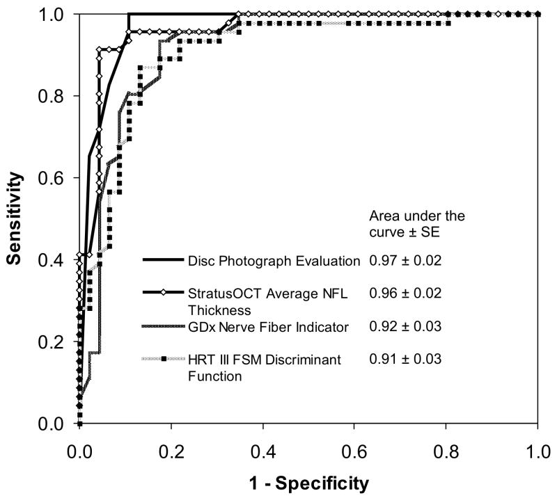
Results: The average (+/- standard deviation) visual field mean deviation for glaucomatous eyes was -4.0 +/- 2.5 dB (decibels). Parameters with largest AUCs (+/- standard error) were: average RNFL thickness for StratusOCT (0.96 +/- 0.02), nerve fiber indicator for GDx VCC (0.92 +/- 0.03), Frederick S. Mikelberg (FSM) discriminant function for HRT III (0.91 +/- 0.03), and 0.97 +/- 0.02 for disk photograph evaluation. At 95% specificity, sensitivity of disk photograph evaluation (90%) was greater than GDx VCC (P = .05) and HRT III (P = .002) results, but not significantly different than those of StratusOCT (P > .05). The combination of StratusOCT average RNFL thickness and HRT III cup-to-disk area with CART produced a sensitivity of 91% and specificity of 96%.
Conclusions: StratusOCT, GDx VCC, and HRT III performed as well as, but not better than, qualitative evaluation of optic disk stereophotographs for detection of early perimetric glaucoma. The combination of StratusOCT average RNFL thickness and HRT III cup-to-disk area ratio provided a high diagnostic precision.
Figures

References
-
- Sommer A, Katz J, Quigley HA, et al. Clinically detectable nerve fiber atrophy precedes the onset of glaucomatous field loss. Arch Ophthalmol. 1991;109:77–83. - PubMed
-
- Johnson CA, Sample PA, Zangwill LM, et al. Structure and function evaluation (SAFE): II. Comparison of optic disk and visual field characteristics. Am J Ophthalmol. 2003;135:148–54. - PubMed
-
- Tuulonen A, Airaksinen PJ. Initial glaucomatous optic disk and retinal nerve fiber layer abnormalities and their progression. Am J Ophthalmol. 1991;111:485–90. - PubMed
-
- Airaksinen PJ, Drance SM, Douglas GR, Schulzer M, Wijsman K. Visual field and retinal nerve fiber layer comparisons in glaucoma. Arch Ophthalmol. 1985;103:205–7. - PubMed
-
- Kass MA, Heuer DK, Higginbotham EJ, et al. The Ocular Hypertension Treatment Study: a randomized trial determines that topical ocular hypotensive medication delays or prevents the onset of primary open-angle glaucoma. Arch Ophthalmol. 2002;120:701–13. discussion 829–30. - PubMed
Publication types
MeSH terms
Grants and funding
LinkOut - more resources
Full Text Sources
Other Literature Sources
Medical
Miscellaneous
