. 2007 Nov 1;311(1):40-52.
doi: 10.1016/j.ydbio.2007.08.010. Epub 2007 Aug 10.

Actomyosin contractility and microtubules drive apical constriction in Xenopus bottle cells

Affiliations

Actomyosin contractility and microtubules drive apical constriction in Xenopus bottle cells

Jen-Yi Lee et al. Dev Biol. .

Abstract

Cell shape changes are critical for morphogenetic events such as gastrulation, neurulation, and organogenesis. However, the cell biology driving cell shape changes is poorly understood, especially in vertebrates. The beginning of Xenopus laevis gastrulation is marked by the apical constriction of bottle cells in the dorsal marginal zone, which bends the tissue and creates a crevice at the blastopore lip. We found that bottle cells contribute significantly to gastrulation, as their shape change can generate the force required for initial blastopore formation. As actin and myosin are often implicated in contraction, we examined their localization and function in bottle cells. F-actin and activated myosin accumulate apically in bottle cells, and actin and myosin inhibitors either prevent or severely perturb bottle cell formation, showing that actomyosin contractility is required for apical constriction. Microtubules were localized in apicobasally directed arrays in bottle cells, emanating from the apical surface. Surprisingly, apical constriction was inhibited in the presence of nocodazole but not taxol, suggesting that intact, but not dynamic, microtubules are required for apical constriction. Our results indicate that actomyosin contractility is required for bottle cell morphogenesis and further suggest a novel and unpredicted role for microtubules during apical constriction.


Figures
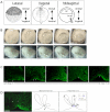
Fig. 1
Fig. 1
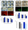
Xenopus laevis bottle cell formation. (A) Embryo Orientation. Lateral illustration (left panel, Stage 8) and vegetal illustration (middle, Stage 10) from Nieuwkoop and Faber series (Nieuwkoop and Faber, 1994). Dotted line through vegetal view shows the mid-sagittal plane. Abbreviations: DMZ, Dorsal Marginal Zone; BC, blastocoel; Br.c, Brachet’s cleft; BP, blastopore. (B) Time-lapse images of bottle cell formation in a whole embryo (top) and in a dorsal-lateral marginal zone explant. Time elapsed (in minutes) noted in the bottom left hand corner of each panel. Small arrows point to sites of bottle cell formation. Movies of this embryo and explant can be found in the supplementary materials. (C) Confocal midsagittal images of DMZs stained with α-tubulin antibody from late stage 9 (left) to stage 10.25 (right) showing progression of bottle cell formation (small arrows) and blastopore groove formation. In this and all DMZ midsagittal views, embryos are oriented with vegetal to the left and dorsal side down. (D) Measuring apical constriction. Left panel shows a confocal image of a midsagittal section of a DMZ stained with α-tubulin antibody. Asterisks indicate cells undergoing apical constriction, which are illustrated in middle and right panels. Blastopore depth (d), indicated by the length of the arrow in the middle panel. The right panel illustrates the Apical Index (A.I.), which is the cell length (l) divided by the apical width (aw). Scale bar = 50 μm.
Fig. 2
Fig. 2
Significant F-actin and activated myosin accumulation occurs at the apical surface of bottle cells. (A) Confocal, midsagittal sections of DMZs stained for F-actin (Oregon green-conjugated phalloidin), activated myosin (anti-pMLC), α-tubulin (anti-DM1α), and memGFP (anti-GFP). (B) Quantification of pixel intensity at the apical surface of bottle cells and non-bottle cells. Asterisks above bottle cell bars indicate p-value < 0.05 compared to non-bottle cells of same staining. Error bars = 2X standard error (s.e.). For morphometrics of cells analyzed, see Supplementary Fig. 3. The following numbers of cells were measured for each subgroup (bottle cells and non-bottle cells number were equivalent): F-actin, 66; pMLC, 66; tubulin, 34; memGFP, 54. (C) Confocal, midsagittal sections of DMZ’s stained for F-actin (Oregon green-conjugated phalloidin). Arrowhead points to marginal zone, and asterisks indicate cells that are accumulating F-actin at their apical membranes without significant cell shape changes. Panels from left (Lateral MZ) to right (Dorsal MZ) represent transition from non-bottle cells to early bottle cells. Scale bar = 50 μm.
Fig. 3
Fig. 3
F-actin is required for bottle cell formation. (A) Actin inhibitors latrunculin B (top) and cytochalasin D (bottom) reversibly prevent bottle cell formation. Control embryos were in 1% DMSO. Bar graph shows percent of embryos making bottle cells in the presence of DMSO control or inhibitor. n, number of embryos. Error bars = 2X s.e. (B) F-actin distribution as indicated by Oregon green-conjugated phalloidin in control, latrunculin B, and cytochalasin D-treated embryos.
Fig. 4
Fig. 4
Myosin function is required for blastopore groove formation and efficient constriction. (A) Blebbistatin-treated whole embryos (top right) and explants (middle right) make bottle cells but exhibit weak apical constriction compared to control (left panels). Blebbistatin-treated embryos have smaller apical indices than control embryos. Bottom row of pictures show control and blebbistatin-treated embryos immunostained with pMLC primary antibody and Texas Red secondary antibody. The Texas Red secondary antibody results in nonspecific staining, allowing visualization of cell outlines for quantitative analysis (compare with Alexa 488 secondary shown in Figs. 2A, 4C, and Supplementary Fig. 2A,B; see Materials and Methods). (B) Apical width, cell length, and apical index in control versus blebbistatin treated bottle cells. Asterisks denote p ≤ 0.001. (C) ML-7 treatment results in shallower blastopore invagination (top) and reduced pMLC staining. (D) Apical width, cell length, and apical index in control versus ML-7 treated bottle cells. (E) Blebbistatin and ML-7 treatment both result in significantly shallower blastopore depths compared to controls. Error bars = 2X s.e.; p ≤ 0.001 for both control vs. blebbistatin and control vs. ML-7. Scale bar = 50 μm.
Fig 5
Fig 5
Intact microtubules are required for efficient apical constriction, but not elongation, of bottle cells. (A) Nocodazole affects bottle cell formation by affecting apical constriction. Middle panels show morphological differences between the bottle cells forming in control versus in nocodazole-treated embryos. Nocodazole treatment disrupts α-tubulin staining. Bar graph shows quantitation of bottle cell morphology. Only apical width and apical index are significantly different in presence of nocodazole (p ≤ 0.0001). (B) Taxol stabilizes microtubules (see also Supplementary Fig. 6) without affecting blastopore formation or bottle cell morphology. Error bars = 2X s.e.
Fig. 6
Fig. 6
Nocodazole treatment does not disrupt F-actin accumulation or MLC phosphorylation. Embryos were cultured in 1% DMSO control (A, C, E) or 15 μg/ml nocodazole (B, D, F), then fixed and stained with anti-α-tubulin (A, B), phalloidin (C, D), or anti-pMLC (E, F). Small arrows point to center of marginal zone (presumptive blastopore).
Fig. 7
Fig. 7
Summary and model of the cytoskeletal mechanisms of Xenopus bottle cell formation. DMZ cells at stage 9 (left) are cuboidal. In unperturbed bottle cells, F-actin (fuchsia) and myosin (orange) are apically localized and intact microtubules (blue) emanate from the apical side, and bottle cells undergo apical constriction and apicobasal elongation while blastopore depth increases as one result of cell shape changes (top row). When F-actin dynamics are inhibited (second row), F-actin does not accumulate apically while pMLC localization is undisturbed (data not shown). Bottle cells do not apically constrict without F-actin, nor do they invaginate to increase blastopore depth. In the presence of myosin inhibitors (third row), F-actin localization still occurs, while pMLC localizes apically in blebbistatin but is reduced in ML-7 treatment. All aspects of bottle cell formation and blastopore depth are disturbed. Nocodazole treatment (bottom row) does not affect F-actin or pMLC localization. Bottle cells without intact microtubules undergo apicobasal elongation normally, but do not apically constrict efficiently, nor do they exhibit significant blastopore depths compared to untreated embryos. Plus signs mean the protein is functional (left columns) or that the event occurs normally (right columns). Minus sign indicates the activity or structure of the protein has been perturbed with inhibitors. Down arrows signify a reduction in cell shape change or decrease in blastopore depth. Question marks indicate unknown results, as those experiments or analyses were not performed. MT, microtubules.

Similar articles

Cited by

References

    1. Baker PC. Fine Structure and Morphogenic Movements in the Gastrula of the Treefrog, Hyla regilla. J Cell Biol. 1965;24:95–116. - PMC - PubMed
    1. Barrett K, Leptin M, Settleman J. The Rho GTPase and a putative RhoGEF mediate a signaling pathway for the cell shape changes in Drosophila gastrulation. Cell. 1997;91:905–15. - PubMed
    1. Benink HA, Bement WM. Concentric zones of active RhoA and Cdc42 around single cell wounds. J Cell Biol. 2005;168:429–39. - PMC - PubMed
    1. Bresnick AR. Molecular mechanisms of nonmuscle myosin-II regulation. Curr Opin Cell Biol. 1999;11:26–33. - PubMed
    1. Burnside B. Microtubules and microfilaments in newt neuralation. Dev Biol. 1971;26:416–41. - PubMed

Publication types