Review
. 2007 Aug;27(3):457-75.
doi: 10.1016/j.iac.2007.07.001.

Cardiovascular manifestations of hypereosinophilic syndromes

Affiliations
Review

Cardiovascular manifestations of hypereosinophilic syndromes

Princess U Ogbogu et al. Immunol Allergy Clin North Am. 2007 Aug.

Abstract

The hypereosinophilic syndromes (HESs) are characterized by persistent marked eosinophilia (>1500 eosinophils/mm(3)), the absence of a primary cause of eosinophilia (such as parasitic or allergic disease), and evidence of eosinophil-mediated end organ damage. Cardiovascular complications of HES are a major source of morbidity and mortality in these disorders. The most characteristic cardiovascular abnormality in HES is endomyocardial fibrosis. Patients who have an HES also may develop thrombosis, particularly in the cardiac ventricles, but also occasionally in deep veins. Because of the rarity of these disorders, specific guidelines for the management of the cardiac and thrombotic complications of HES are lacking. This article reviews the diagnosis and management of the cardiovascular manifestations of HES.


Figures

Fig. 1
Fig. 1
Fatal endomyocardial fibrosis in a patient with HES and FIP1L1/PGDFRA mutation. (From Stone RM, Gilliland DG, Klion AD. Platelet-derived growth factor receptor inhibition to treat idiopathic hypereosinophilic syndrome. Semin Oncol. Apr 2004;31(2 Suppl 6):12-17 with permission.)
Fig. 2
Fig. 2
Echocardiogram of the same patient presented in Fig 1 . A) In the parasternal long axis view, an area of increased echo density (arrow) denotes thrombus and/or fibrosis along the basal posterior wall of the left ventricle which also involves the posterior leaflet of the mitral valve. B) Color Doppler in the same view demonstrates significant mitral regurgitation. C) In the short axis view shown during diastole, there is extensive thrombus and/or fibrosis on the posterior wall (arrow) with compromise of the left ventricular cavity size. (Courtesy of Dr. Vandana Sachdev, National Heart, Lung, and Blood Institute)
Fig. 2
Fig. 2
Echocardiogram of the same patient presented in Fig 1 . A) In the parasternal long axis view, an area of increased echo density (arrow) denotes thrombus and/or fibrosis along the basal posterior wall of the left ventricle which also involves the posterior leaflet of the mitral valve. B) Color Doppler in the same view demonstrates significant mitral regurgitation. C) In the short axis view shown during diastole, there is extensive thrombus and/or fibrosis on the posterior wall (arrow) with compromise of the left ventricular cavity size. (Courtesy of Dr. Vandana Sachdev, National Heart, Lung, and Blood Institute)
Fig. 3
Fig. 3
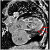
Cine MRI in a 4-chamber view of the same patient presented in Fig. 1 using a steady-state free precession technique. Note that the right ventricular cavity is nearly obliterated. Abbreviations: LV = left ventricle, RV = right ventricle, RA = right atrium, LA = left atrium. (Courtesy of Drs. Patricia Bandettini and Andrew Arai, National Heart, Lung, and Blood Institute)
Fig. 4
Fig. 4
Delayed hyper-enhancement images of the same patient presented in Fig. 1 obtained 20 minutes after administration of gadolinium contrast using an inversion recovery gradient echo technique validated for imaging myocardial infarction. There is a bright white rim of enhancement lining the left ventricle (arrows) which is consistent with endomyocardial fibrosis. (Courtesy of Drs. Patricia Bandettini and Andrew Arai, National Heart, Lung, and Blood Institute)
Fig. 5
Fig. 5
Histology of fatal endomyocardial fibrosis in the same patient presented in Fig. 1. Arrow denotes areas of intracardiac fibrosis bordered by areas of unaffected myocardium.

Similar articles

Cited by

References

    1. Chusid MJ, Dale DC, West BC, Wolff SM. The hypereosinophilic syndrome: analysis of fourteen cases with review of the literature. Medicine (Baltimore) 1975 Jan;54(1):1–27. - PubMed
    1. Ommen SR, Seward JB, Tajik AJ. Clinical and echocardiographic features of hypereosinophilic syndromes. Am J Cardiol. 2000 Jul 1;86(1):110–113. - PubMed
    1. Parrillo JE, Borer JS, Henry WL, Wolff SM, Fauci AS. The cardiovascular manifestations of the hypereosinophilic syndrome. Prospective study of 26 patients, with review of the literature. Am J Med. 1979 Oct;67(4):572–582. - PubMed
    1. Ginsberg F, Parrillo JE. Eosinophilic myocarditis. Heart Fail Clin. 2005 Oct;1(3):419–429. - PubMed
    1. Cooper LT, Zehr KJ. Biventricular assist device placement and immunosuppression as therapy for necrotizing eosinophilic myocarditis. Nat Clin Pract Cardiovasc Med. 2005 Oct;2(10):544–548. - PubMed

Publication types

MeSH terms