. 2007 Nov 15;311(2):636-49.
doi: 10.1016/j.ydbio.2007.08.015. Epub 2007 Aug 16.

The GATA factor Serpent cross-regulates lozenge and u-shaped expression during Drosophila blood cell development

Affiliations

The GATA factor Serpent cross-regulates lozenge and u-shaped expression during Drosophila blood cell development

Selen Muratoglu et al. Dev Biol. .

Abstract

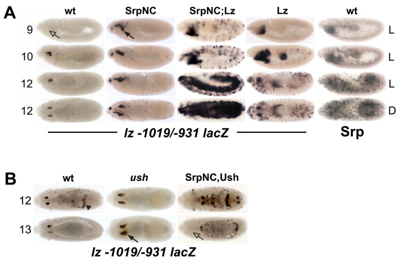
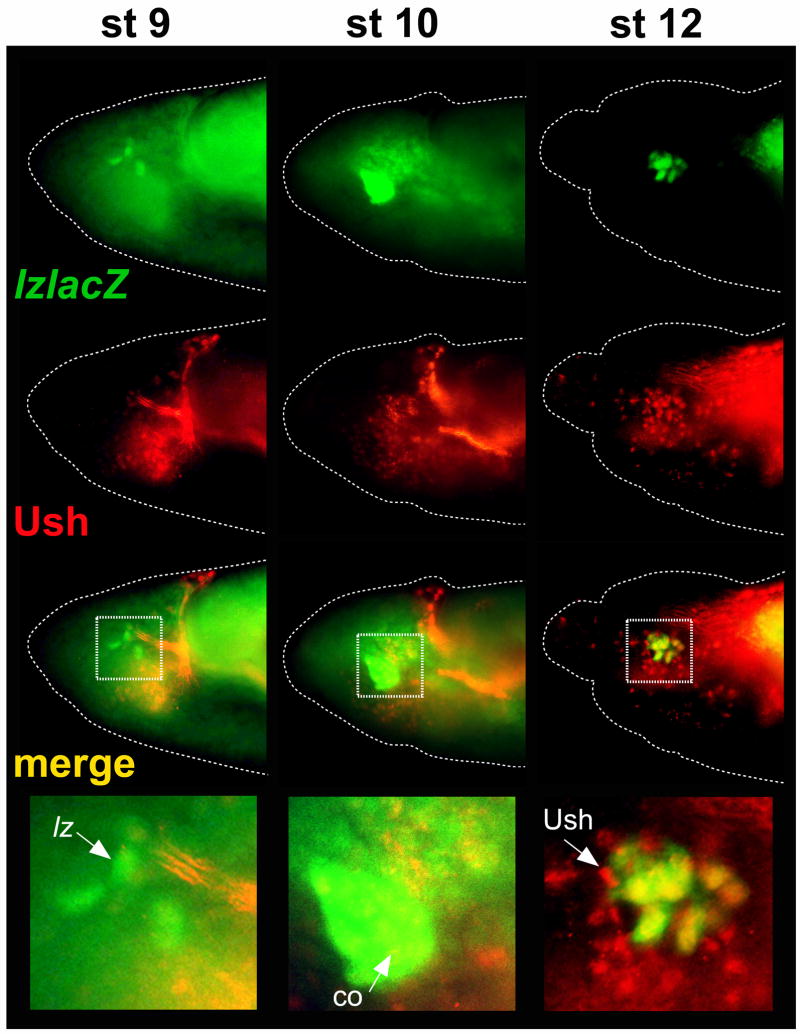
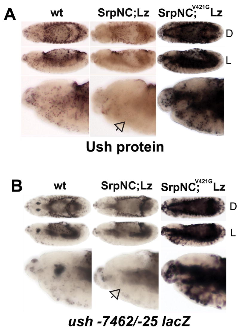
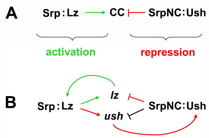
The Drosophila GATA factor Serpent interacts with the RUNX factor Lozenge to activate the crystal cell program, whereas SerpentNC binds the Friend of GATA protein U-shaped to limit crystal cell production. Here, we identified a lozenge minimal hematopoietic cis-regulatory module and showed that lozenge-lacZ reporter-gene expression was autoregulated by Serpent and Lozenge. We also showed that upregulation of u-shaped was delayed until after lozenge activation, consistent with our previous results that showed u-shaped expression in the crystal cell lineage is dependent on both Serpent and Lozenge. Together, these observations describe a feed forward regulatory motif, which controls the temporal expression of u-shaped. Finally, we showed that lozenge reporter-gene activity increased in a u-shaped mutant background and that forced expression of SerpentNC with U-shaped blocked lozenge- and u-shaped-lacZ reporter-gene activity. This is the first demonstration of GATA:FOG regulation of Runx and Fog gene expression. Moreover, these results identify components of a Serpent cross-regulatory sub-circuit that can modulate lozenge expression. Based on the sub-circuit design and the combinatorial control of crystal cell production, we present a model for the specification of a dynamic bi-potential regulatory state that contributes to the selection between a Lozenge-positive and Lozenge-negative state.


Figures

Fig. 1
Fig. 1. Identification of the lz crystal cell cis-regulatory module
(A) Schematic of the lz locus and screen for the crystal cell CRM. A horizontal arrow marks the transcriptional start site, designated position 0. Solid lines indicate introns, whereas boxes indicate exons. The upstream c11.1 gene is depicted in grey. Beneath the 21 kb map is an expanded map of the 1.5 kb region upstream of the lz transcription unit. Evolutionarily conserved regions are highlighted in yellow. The relative position of the P-element pGawB insert is indicated. The cryptic promoter used by this transgene likely lies just 5′ to the transcription start site. The DNA fragments used to screen for crystal cell CRMs are indicated by black lines and are positioned and numbered relative to the transcription start site. Solid lines represent fragments with crystal cell activity, whereas dashed lines represent fragments lacking crystal cell activity. The red diamonds designate representative data presented in part B. (B) Fragment-driven reporter-gene (lacZ) expression during embryogenesis and in third larval instar lymph gland. The stage of embryogenesis is indicated at the left of each row. The lz-lacZ strain designations are indicated at the top of each column. Orientation of the embryo is indicated to the right of each row. Solid arrows mark activity and open arrows mark the lack of activity in stage 8 hemocyte precursors and stage 9 crystal cell precursors. The boundaries between the cortical and medullary zones of the larval lymph glands are outlined. Abbreviations: lz, lozenge; llg, third larval instar lymph gland; cz, cortical zone; mz, medullary zone; L, lateral view; D, dorsal view.
Fig. 2
Fig. 2. Clustered GATA and RUNX motifs are required for lz crystal cell CRM activity
(A) Schematic showing fragments with mutated GATA and RUNX motifs used to evaluate lz-lacZ CRM activity. The 1.5 kb region upstream of the lz transcription start site is depicted with the evolutionarily conserved regions highlighted in yellow. Beneath the map of the 1.5 kb region is an expanded map of the region from −1250 to −727. Red vertical lines show the relative positions of the 13 GATA motifs between positions −1207 and −768. The blue diamonds mark the positions of the two RUNX motifs located at −995 and −977. Mutations in the four GATA motifs, between positions −1010 and −947, are designated G4m, whereas the mutations in the two RUNX motifs are designated R2m. The mutated fragments are numbered and positioned relative to the transcription start site, with the suffix G4m or R2m to indicate disruption of the GATA or RUNX motifs, respectively. Solid line represents a fragment with crystal cell activity, whereas dashed lines represent fragments lacking crystal cell activity. (B) Comparison of wild-type and GATA or RUNX mutant CRMs. Dorsal views of stage 13 embryos. lz-lacZ strains are indicated in the lower right hand corner of each panel. Closed arrows mark activity in crystal cells; open arrows mark lack of activity. Abbreviations: G4m, GATA Core mutants; R2m, RUNX mutants.
Fig. 3
Fig. 3. SrpNC is a positive and negative regulator of lz crystal cell CRM activity
(A) SrpNC and Lz synergistically upregulate lz −1019/−931 lacZ activity. From left to right: embryos in the first four columns are stained with α-β-galactosidase antibody; embryos in the last column are stained with α-Srp antibody. (B) SrpNC and Ush block lz −1019/−931 lacZ activity. lz −1019/−931 lacZ activity was assessed in different genetic backgrounds and developmental stages. The stage of embryogenesis is indicated at the left of each row. The genetic background is indicated at the top of each column. Orientation of the embryo is indicated at the right of each row (panel A only). SrpNC indicates twi-Gal4 driving UAS-srpNC. Lz indicates twi-Gal4 driving UAS-lz. SrpNC;Lz represents twi-Gal4 driving UAS-srpNC and UAS-lz located on chromosomes II and III, respectively. SrpNC,Ush represents twi-Gal4 driving UAS-srpNC and UAS-ush located on chromosome II. Closed arrows mark increased activity in crystal cells; open arrow marks lack of activity; arrowhead marks ectopic expression in the amnioserosa, which is used to identify embryos carrying the reporter-gene. Abbreviations: wt, wild-type; Srp, Serpent; Lz, Lozenge; Ush, U-shaped; L, lateral view; D, dorsal view.
Fig. 4
Fig. 4. lz-lacZ and Ush expression during crystal cell development
The lz −1236/−737 CRM was used to assess lz expression and is depicted in green. Ush protein expression is depicted in red. Co-localized expression is depicted in yellow. The stage of embryogenesis is indicated at the top of each column. The enlarged area is marked by a white dotted box. Arrows mark the following: stage 9 cells expressing only lz-lacZ; stage 10 cells with co-localized expression of lz-lacZ and Ush; and stage 12 cells expressing only Ush. Abbreviations: lz-lacZ, lz −1236/−747 lacZ; Ush, U-shaped; co, co-localization.
Fig. 5
Fig. 5. N-terminal zinc-finger conserved valine residue is required for SrpNC and Lz repression of ush expression
(A) Ush protein expression and (B) ush −7462/−25 lacZ activity was assessed in stage 13 embryos with different genetic backgrounds. The genetic background is indicated at the top of each column. Orientation of the embryo is indicated at the right of each row. SrpNC;Lz represents twi-Gal4 driving UAS-srpNC and UAS-lz located on chromosomes II and III, respectively. SrpNC;V421G Lz represents twi-Gal4 driving UAS-srpNCV4216 and UAS-lz located on chromosomes II and III, respectively. Open arrows mark lack of expression in hemocytes. Abbreviations: wt, wild-type; Srp, Serpent; Lz, Lozenge; Ush, U-shaped; L, lateral view; D, dorsal view.
Fig. 6
Fig. 6. Ush competes with Lz to control SrpNC regulation of ush expression
(A) ush −7462/−25 lacZ and (B) ush −174/−25 lacZ was assessed in different genetic backgrounds and developmental stages. The stage of embryogenesis is indicated at the left of each row. All embryos are lateral views. The genetic background is indicated at the top of each column. SrpNC;Lz represents twi-Gal4 driving UAS-srpNC and UAS-lz located on chromosome II and III, respectively. SrpNC,Ush represents twi-Gal4 driving UAS-srpNC and UAS-ush located on chromosome II. SrpNC,Ush;Lz indicates twi-Gal4 driving UAS-srpNC,UAS-ush and UAS-lz located on chromosomes II and III, respectively. Plasmatocytes and crystal cells (outlined in panel A) are marked with arrows: closed arrows indicate wild-type CRM activity; open arrows indicate reduced activity; open dotted arrows indicate lack of activity. The level of CRM activity in the head mesoderm is marked with arrowheads: closed arrowheads indicate increased activity; the open arrowhead indicates lack of activity. Abbreviations: wt, wild-type; Srp, Serpent; Lz, Lozenge; Ush, U-shaped; cc, crystal cells; pl, plasmatocytes.
Fig. 7
Fig. 7. Model of Srp cross-regulatory control of crystal cell lineage commitment
(A) Combinatorial control of crystal cell lineage commitment by Srp, Lz, and Ush. Srp acts as a contextual switch, interacting with Lz to activate crystal cell lineage commitment and with Ush to block crystal cell production. (B) Srp cross-regulatory control of lz and ush expression. Srp and Lz interact to maintain the Lz-positive regulatory state. These transcriptional regulators also activate ush expression in Lz-positive crystal cell precursors to produce a dynamic, bi-potential regulatory state. SrpNC mediates cross-antagonism of Lz and Ush, acting with Lz to maintain the Lz-positive state or with Ush to select the Lz-negative regulatory state. Additionally, the SrpNC:Ush complex can downregulate ush expression, thereby limiting the capacity of the complex to block lz expression. Srp cross-regulatory control of lz and ush is mediated through the GATA and RUNX binding sites within the minimal hematopoietic CRMs of each locus. Green arrows indicate the activation pathway. Red arrows and blocked red lines indicate the repression pathway. The blocked black line indicates relief of repression. Abbreviations: Srp, Serpent; Lz, Lozenge, CC, crystal cells; Ush, U-shaped.

Similar articles

Cited by

References

    1. Alfonso TB, Jones BW. gcm2 promotes glial cell differentiation and is required with glial cells missing for macrophage development in Drosophila. Dev Biol. 2002;248:369–383. - PubMed
    1. Anglin I, Passaniti A. Runx protein signaling in human cancers. Cancer Treat Res. 2004;119:189–215. - PubMed
    1. Bataille L, Auge B, Ferjoux G, Haenlin M, Waltzer L. Resolving embryonic blood cell fate choice in Drosophila: interplay of GCM and RUNX factors. Development. 2005;132:4635–4644. - PubMed
    1. Bernardoni R, Vivancos V, Giangrande A. glide/gcm is expressed and required in the scavenger cell lineage. Dev Biol. 1997;191:118–130. - PubMed
    1. Brand AH, Perrimon N. Targeted gene expression as a means of altering cell fates and generating dominant phenotypes. Development. 1993;118:401–415. - PubMed

Publication types

MeSH terms

LinkOut - more resources