

doi: 10.1016/j.febslet.2007.08.076.
Epub 2007 Sep 7.
Extensive mutagenesis experiments corroborate a structural model for the DNA deaminase domain of APOBEC3G
Affiliations
- PMID: 17869248
- PMCID: PMC2014798
- DOI: 10.1016/j.febslet.2007.08.076
Item in Clipboard
Extensive mutagenesis experiments corroborate a structural model for the DNA deaminase domain of APOBEC3G
FEBS Lett.
.
Abstract
APOBEC3G is a single-strand DNA cytosine deaminase capable of blocking retrovirus and retrotransposon replication. APOBEC3G has two conserved zinc-coordinating motifs but only one is required for catalysis. Here, deletion analyses revealed that the minimal catalytic domain consists of residues 198-384. Size exclusion assays indicated that this protein is monomeric. Many (31/69) alanine substitution derivatives of APOBEC3G198-384 retained significant to full levels of activity. These data corroborated an APOBEC2-based structural model for the catalytic domain of APOBEC3G indicating that most non-essential residues are solvent accessible and most essential residues cluster within the protein core.
Figures

APOBEC3G deletion mutants delineate a minimal active domain. (A) An illustration showing the amino acid boundaries used for deletion constructs. The HXE-X23–28-CX2–4C motifs are depicted by open boxes, and the asterisk designates the catalytic domain. (B) RifR mutation frequencies of the indicated GST-A3G constructs. Each X represents the mutation frequency of an individual culture (n=8 per construct). The median mutation frequency for cells expressing the vector control, A3G, A3G175-384 and A3G198-384 is indicated. (C) Size exclusion profiles of GST (25 kDa), A3G198-384 (22 kDa) and lysozyme (14 kDa) indicate that A3G198-384 is monomeric.
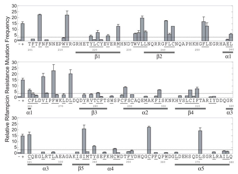
Mutator phenotype of 69 APOBEC3G alanine substitution mutants. Histograms showing the relative RifR mutation frequencies of cells expressing the vector control (−), A3G198-384 (+) or derivatives with alanine substitutions at the underlined amino acid positions. Each histogram bar reports data from 2–5 independent experiments, except the bars for vector and A3G198-384, which reflect data from 12 (± SEM). The dotted line represents the 3-fold significance threshold that was used to distinguish active and inactive mutant constructs. A3G amino acids 198-200 and 381-384 were not shown. A predicted secondary structure of A3G198-384, based on a pair wise alignment with APOBEC2, is included to facilitate comparisons with Fig. 4.
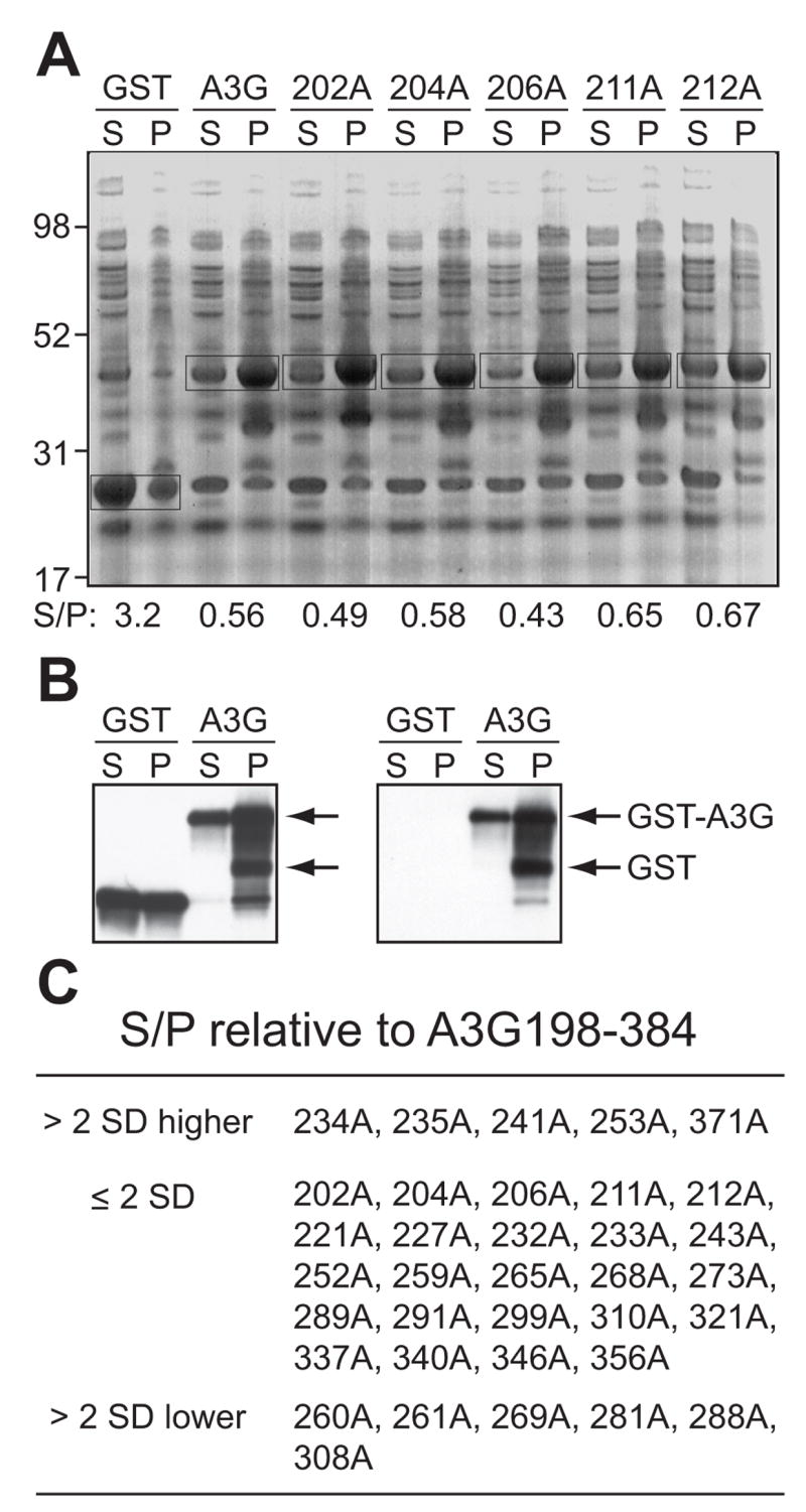
GST-A3G198-384 expression data. (A) A representative gel showing the soluble (supernatant) and insoluble (pellet) amounts of GST, GST-A3G198-384 (WT) and 5 mutant derivatives. The S/P ratio of the boxed bands is shown below each lane. The E. coli protein(s) that migrates indistinguishably from GST-A3G198-384 was present in every sample and it provided a constant (but lower) background signal. (B) Anti-GST (left panel) and anti-A3G (right panel) immunoblots confirm the identities of the bands boxed in Fig. 3A. (C) Mutants with higher, near equivalent or lower S/P values than those of wild type GST-A3G198-384 are listed. Total protein amounts (S+P) were similar for every construct ([A] and data not shown).
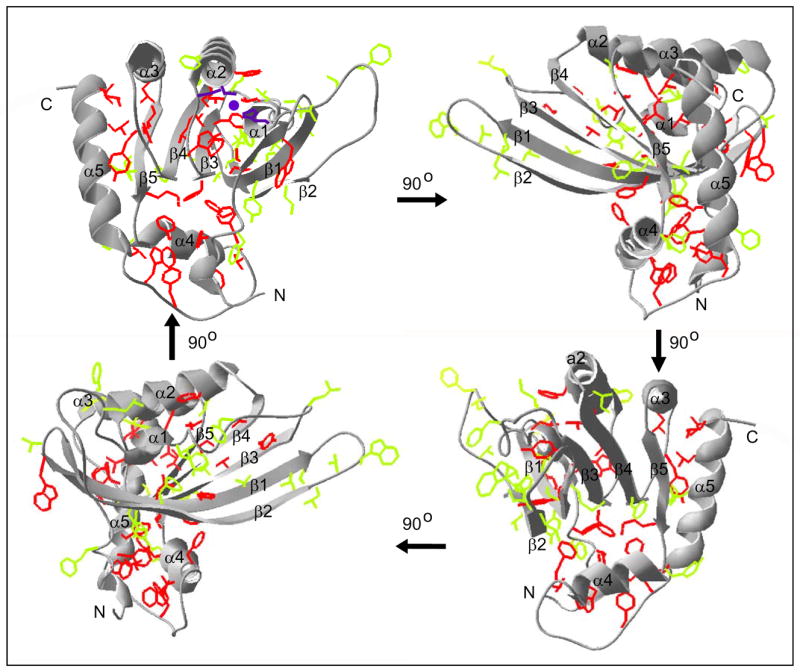
Four views of a model A3G198-384 structure based on human APOBEC2 [16]. The predicted α helices and β sheets correspond with those shown in Fig. 2. Relevant amino acid side chains are depicted in green or red to reflect the activity or inactivity, respectively, of an alanine substitution at that position. The purple amino acid side chains in the top left panel represent the zinc (purple ball)-coordinating histidine (H257 in α1) and cysteine residues (C288 and C291 in α2).
References
-
- Longerich S, Basu U, Alt F, Storb U. AID in somatic hypermutation and class switch recombination. Curr Opin Immunol. 2006;18:164–74. - PubMed
-
- Sheehy AM, Gaddis NC, Choi JD, Malim MH. Isolation of a human gene that inhibits HIV-1 infection and is suppressed by the viral Vif protein. Nature. 2002;418:646–50. - PubMed
-
- Harris RS, Petersen-Mahrt SK, Neuberger MS. RNA editing enzyme APOBEC1 and some of its homologs can act as DNA mutators. Molecular Cell. 2002;10:1247–53. - PubMed
-
- Harris RS, Bishop KN, Sheehy AM, Craig HM, Petersen-Mahrt SK, Watt IN, Neuberger MS, Malim MH. DNA deamination mediates innate immunity to retroviral infection. Cell. 2003;113:803–9. - PubMed
Publication types
MeSH terms
Substances
Grants and funding
LinkOut - more resources
Full Text Sources
Other Literature Sources
