

Differential effects of aging and Alzheimer's disease on medial temporal lobe cortical thickness and surface area
- PMID: 17869384
- PMCID: PMC3703585
- DOI: 10.1016/j.neurobiolaging.2007.07.022
Differential effects of aging and Alzheimer's disease on medial temporal lobe cortical thickness and surface area
Abstract
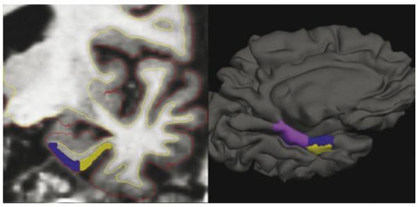
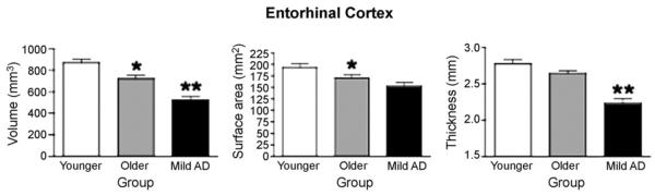
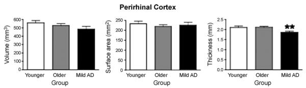
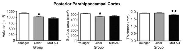
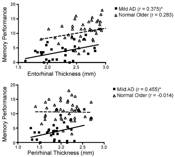
The volume of parcellated cortical regions is a composite measure related to both thickness and surface area. It is not clear whether volumetric decreases in medial temporal lobe (MTL) cortical regions in aging and Alzheimer's disease (AD) are due to thinning, loss of surface area, or both, nor is it clear whether aging and AD differ in their effects on these properties. Participants included 28 Younger Normals, 47 Older Normals, and 29 patients with mild AD. T1-weighted MRI data were analyzed using a novel semi-automated protocol (presented in a companion article) to delineate the boundaries of entorhinal (ERC), perirhinal (PRC), and posterior parahippocampal (PPHC) cortical regions and calculate their mean thickness, surface area, and volume. Compared to Younger Normals, Older Normals demonstrated moderately reduced ERC and PPHC volumes, which were due primarily to reduced surface area. In contrast, the expected AD-related reduction in ERC volume was produced by a large reduction in thickness with minimal additional effect (beyond that of aging) on surface area. PRC and PPHC also showed large AD-related reductions in thickness. Of all these MTL morphometric measures, ERC and PRC thinning were the best predictors of poorer episodic memory performance in AD. Although the volumes of MTL cortical regions may decrease with both aging and AD, thickness is relatively preserved in normal aging, while even in its mild clinical stage, AD is associated with a large degree of thinning of MTL cortex. These differential morphometric effects of aging and AD may reflect distinct biologic processes and ultimately may provide insights into the anatomic substrates of change in memory-related functions of MTL cortex.
Figures
References
-
- Amunts K, Kedo O, Kindler M, Pieperhoff P, Mohlberg H, Shah NJ, Habel U, Schneider F, Zilles K. Cytoarchitectonic mapping of the human amygdala, hippocampal region and entorhinal cortex: intersubject variability and probability maps. Anat. Embryol. (Berl.) 2005;210:343–352. - PubMed
-
- Bailey P, von Bonin G. The Isocortex of Man. University of Illinois Press; Urbana, IL: 1951.
-
- Berg L, McKeel DW, Jr., Miller JP, Storandt M, Rubin EH, Morris JC, Baty J, Coats M, Norton J, Goate AM, Price JL, Gearing M, Mirra SS, Saunders AM. Clinicopathologic studies in cognitively healthy aging and Alzheimer's disease: relation of histologic markers to dementia severity, age, sex, and apolipoprotein E genotype. Arch. Neurol. 1998;55:326–335. - PubMed
Publication types
MeSH terms
Grants and funding
- U24 RR021382/RR/NCRR NIH HHS/United States
- P01-AG03991/AG/NIA NIH HHS/United States
- P41-RR14075/RR/NCRR NIH HHS/United States
- R01 AG029411/AG/NIA NIH HHS/United States
- R01 NS052585-01/NS/NINDS NIH HHS/United States
- R01 EB001550/EB/NIBIB NIH HHS/United States
- U54 EB005149/EB/NIBIB NIH HHS/United States
- P01 AG003991/AG/NIA NIH HHS/United States
- P50 AG005681/AG/NIA NIH HHS/United States
- R01 NS052585/NS/NINDS NIH HHS/United States
- R01 RR16594-01A1/RR/NCRR NIH HHS/United States
- P50-AG05681/AG/NIA NIH HHS/United States
- U24-RR021382/RR/NCRR NIH HHS/United States
- R21 AG029840/AG/NIA NIH HHS/United States
- R01 RR016594/RR/NCRR NIH HHS/United States
- K23 AG022509/AG/NIA NIH HHS/United States
- K23-AG22509/AG/NIA NIH HHS/United States
LinkOut - more resources
Full Text Sources
Medical
