

Real-time intraoperative ureteral guidance using invisible near-infrared fluorescence
- PMID: 17870110
- PMCID: PMC2505174
- DOI: 10.1016/j.juro.2007.06.049
Real-time intraoperative ureteral guidance using invisible near-infrared fluorescence
Abstract
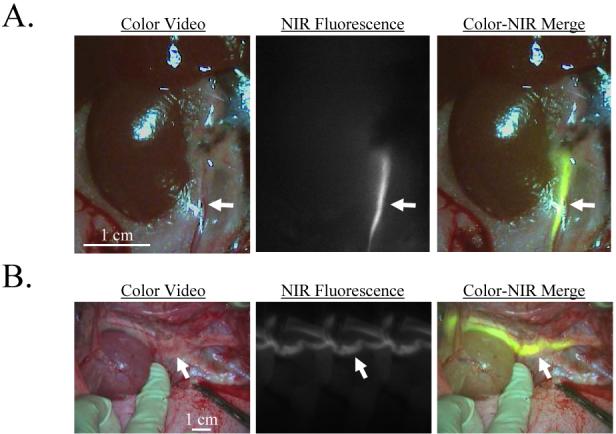
Purpose: Invisible near-infrared light is safe and it penetrates relatively deeply through tissue and blood without altering the surgical field. Our hypothesis was that near-infrared fluorescence imaging would enable visualization of the ureteral anatomy and flow intraoperatively and in real time.
Materials and methods: CW800-CA (LI-COR, Lincoln, Nebraska), the carboxylic acid form of near-infrared fluorophore IRDye 800CW, was injected intravenously, and its renal clearance kinetics and imaging performance were quantified in 350 gm rats and 35 kg pigs. High performance liquid chromatography and electrospray time-of-flight mass spectrometry were used to characterize CW800-CA metabolism in urine. The clinically available near-infrared fluorophore indocyanine green was also used via retrograde injection into the ureter. Using the 2 near-infrared fluorophores the ureters were imaged under the conditions of steady state, intraluminal foreign bodies and injury.
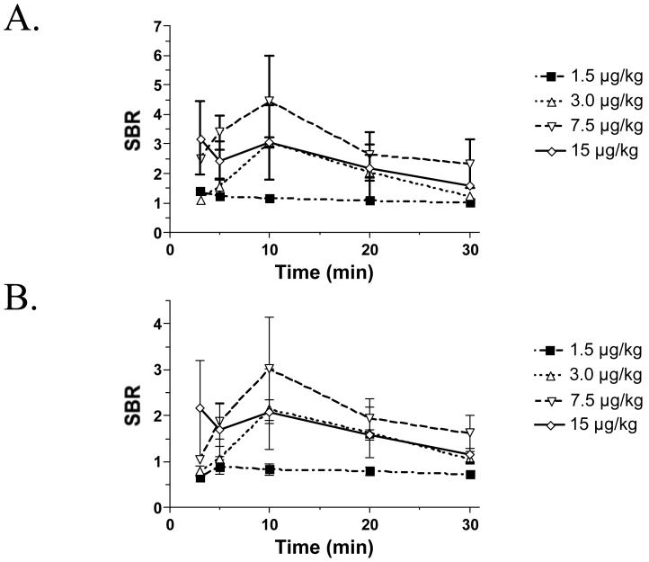
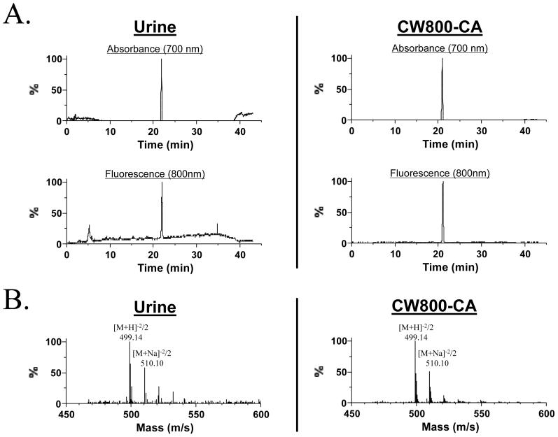
Results: In rat models the highest signal-to-background ratio for visualization occurred after intravenous injection of 7.5 microg/kg CW800-CA with values of 4.0 or greater and 2.3 or greater at 10 and 30 minutes, respectively. In pig models 7.5 microg/kg CW800-CA clearly visualized the normal ureter and intraluminal foreign bodies as small as 2.5 mm in diameter. Retrograde injection of 10 microM indocyanine green also permitted the detection of normal ureter and pinpointed urine leakage caused by injury. Electrospray time-of-flight mass spectrometry, and absorbance and fluorescence spectral analysis confirmed that the fluorescent material in urine was chemically identical to CW800-CA.
Conclusions: A convenient intravenous injection of CW800-CA or direct injection of indocyanine green permits high sensitivity visualization of the ureters under steady state and abnormal conditions using invisible light.
Figures
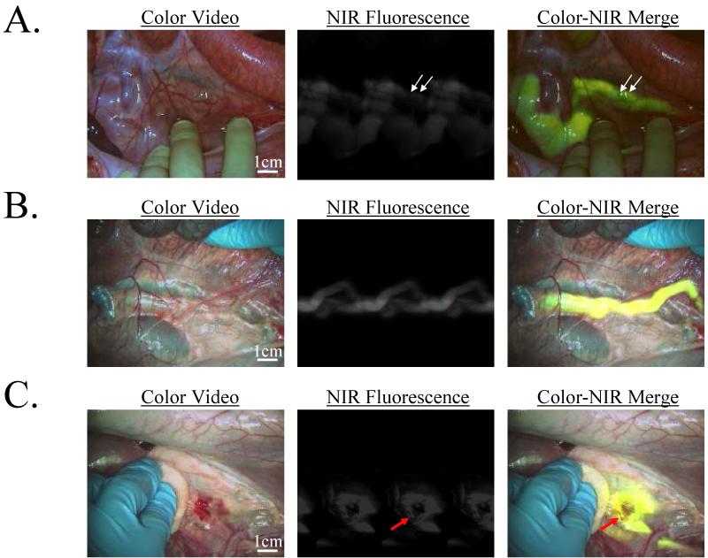
After IV injection of 7.5 μg/kg CW800-CA in the pig, the ureters could be visualized for the next 60 min. Shown is the identification of two foreign bodies (2.5 mm diameter beads; arrows) within the lumen of the ureter. NIR fluorescence exposure times were 100 msec. Data are representative of results from N = 4 independent experiments.
Retrograde injection of 10 ml of 10 μM ICG in saline provided immediate visualization of the ureters. NIR fluorescence exposure times were 60 msec. Data are representative of N = 2 independent experiments.
A site of injury to the ureter made during tissue dissection, in this case from a scalpel, can be pinpointed immediately (red arrow) and repaired. NIR fluorescence exposure times were 60 msec. Data are representative of N = 2 independent experiments.
References
-
- Bothwell WN, Bleicher RJ, Dent TL. Prophylactic ureteral catheterization in colon surgery. A five-year review. Dis Colon Rectum. 1994;37:330. - PubMed
-
- Wood EC, Maher P, Pelosi MA. Routine use of ureteric catheters at laparoscopic hysterectomy may cause unnecessary complications. J Am Assoc Gynecol Laparosc. 1996;3:393. - PubMed
-
- Park S, Pearle MS. Imaging for percutaneous renal access and management of renal calculi. Urol Clin North Am. 2006;33:353. - PubMed
-
- Jabs CF, Drutz HP. The role of intraoperative cystoscopy in prolapse and incontinence surgery. Am J Obstet Gynecol. 2001;185:1368. - PubMed
Publication types
MeSH terms
Substances
Grants and funding
LinkOut - more resources
Full Text Sources
Other Literature Sources
Medical
