

The tooth attachment mechanism defined by structure, chemical composition and mechanical properties of collagen fibers in the periodontium
- PMID: 17870156
- PMCID: PMC2423002
- DOI: 10.1016/j.biomaterials.2007.08.031
The tooth attachment mechanism defined by structure, chemical composition and mechanical properties of collagen fibers in the periodontium
Abstract
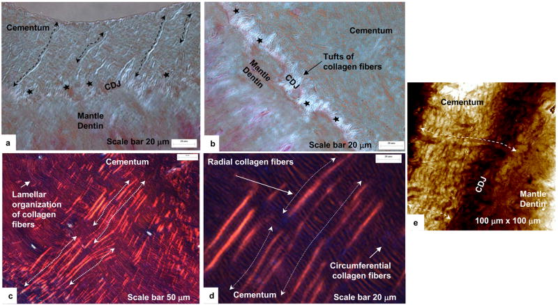
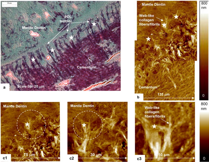
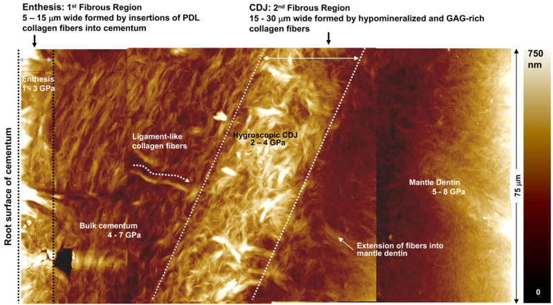
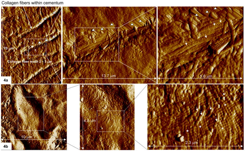
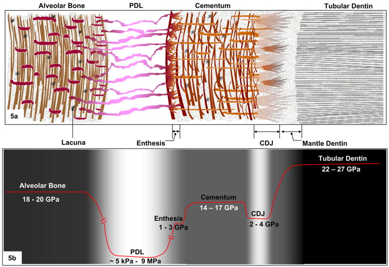
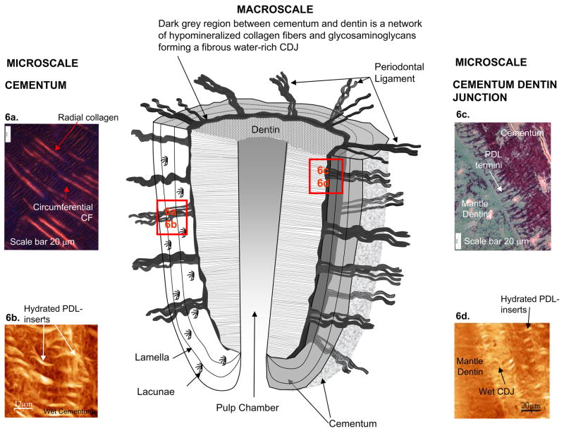
In this study, a comparison between structure, chemical composition and mechanical properties of collagen fibers at three regions within a human periodontium, has enabled us to define a novel tooth attachment mechanism. The three regions include, (1) the enthesis region: insertion site of periodontal ligament (PDL) fibers (collagen fibers) into cementum at the root surface, (2) bulk cementum, and (3) the cementum-dentin junction (CDJ). Structurally, continuity in collagen fibers was observed from the enthesis, through bulk cementum and CDJ. At the CDJ the collagen fibers split into individual collagen fibrils and intermingled with the extracellular matrix of mantle dentin. Under wet conditions, the collagen fibers at the three regions exhibited significant swelling suggesting a composition rich in polyanionic molecules such as glycosaminoglycans. Additionally, site-specific indentation illustrated a comparable elastic modulus between collagen fibers at the enthesis (1-3 GPa) and the CDJ (2-4 GPa). However, the elastic modulus of collagen fibers within bulk cementum was higher (4-7 GPa) suggesting presence of extrafibrillar mineral. It is known that the tooth forms a fibrous joint with the alveolar bone, which is termed a gomphosis. Although narrower in width than the PDL space, the hygroscopic CDJ can also be termed as a gomphosis; a fibrous joint between cementum and root dentin capable of accommodating functional loads similar to that between cementum and alveolar bone. From an engineering perspective, it is proposed that a tooth contains two fibrous joints that accommodate the masticatory cyclic loads. These joints are defined by the attachment of dissimilar materials via graded stiffness interfaces, such as: (1) alveolar bone attached to cementum with the PDL; and (2) cementum to root dentin with the CDJ. Thus, through variations in concentrations of basic constituents, distinct regions with characteristic structures and graded properties allow for attachment and the load bearing characteristics of a tooth.
Figures
Similar articles
-
Ultrastructure and nanomechanical properties of cementum dentin junction.J Biomed Mater Res A. 2004 Feb 1;68(2):343-51. doi: 10.1002/jbm.a.20061. J Biomed Mater Res A. 2004. PMID: 14704976
-
Structure, chemical composition and mechanical properties of human and rat cementum and its interface with root dentin.Acta Biomater. 2009 Feb;5(2):707-18. doi: 10.1016/j.actbio.2008.08.013. Epub 2008 Sep 9. Acta Biomater. 2009. PMID: 18829402 Free PMC article.
-
Adaptive properties of human cementum and cementum dentin junction with age.J Mech Behav Biomed Mater. 2014 Nov;39:184-96. doi: 10.1016/j.jmbbm.2014.07.015. Epub 2014 Jul 24. J Mech Behav Biomed Mater. 2014. PMID: 25133753 Free PMC article.
-
Biomechanical pathways of dentoalveolar fibrous joints in health and disease.Periodontol 2000. 2020 Feb;82(1):238-256. doi: 10.1111/prd.12306. Periodontol 2000. 2020. PMID: 31850635 Review.
-
Developmental pathways of periodontal tissue regeneration: Developmental diversities of tooth morphogenesis do also map capacity of periodontal tissue regeneration?J Periodontal Res. 2019 Feb;54(1):10-26. doi: 10.1111/jre.12596. Epub 2018 Sep 12. J Periodontal Res. 2019. PMID: 30207395 Review.
Cited by
-
Implant Fibrosis and the Underappreciated Role of Myofibroblasts in the Foreign Body Reaction.Cells. 2021 Jul 15;10(7):1794. doi: 10.3390/cells10071794. Cells. 2021. PMID: 34359963 Free PMC article. Review.
-
Dynamic Modelling of Tooth Deformation Using Occlusal Kinematics and Finite Element Analysis.PLoS One. 2016 Mar 31;11(3):e0152663. doi: 10.1371/journal.pone.0152663. eCollection 2016. PLoS One. 2016. PMID: 27031836 Free PMC article.
-
Nanotechnology in Dental Sciences: Moving towards a Finer Way of Doing Dentistry.Materials (Basel). 2010 Mar;3(3):1674-1691. doi: 10.3390/ma3031674. Epub 2010 Mar 8. Materials (Basel). 2010. PMID: 27103959 Free PMC article.
-
Rab27a-mediated extracellular vesicle secretion contributes to osteogenesis in periodontal ligament-bone niche communication.Sci Rep. 2023 May 25;13(1):8479. doi: 10.1038/s41598-023-35172-x. Sci Rep. 2023. PMID: 37231020 Free PMC article.
-
Regulators of Collagen Fibrillogenesis during Molar Development in the Mouse.Front Physiol. 2017 Aug 2;8:554. doi: 10.3389/fphys.2017.00554. eCollection 2017. Front Physiol. 2017. PMID: 28824450 Free PMC article.
References
-
- McIntosh JE, Anderton X, Flores-De-Jacoby L, Carlson DS, Shuler CF, Diekwisch TG. Caiman periodontium as an intermediate between basal vertebrate ankylosis-type attachment and mammalian “true” periodontium. Microsc Res Tech. 2002;59(5):449–59. - PubMed
-
- Ten Cate R. Oral histology: development, structure, and function. Mosby-Year Book Inc.; St. Loius, MI: 1998. p. 236.
-
- Selvig KA. The fine structure of human cementum. Acta Ondontol Scand. 1965;23:423–441. - PubMed
-
- Furseth R. The fine structure of acellular cementum in human young premolars. Scand J Dent Res. 1974;82:437–441. - PubMed
-
- Dewey KW. Normal and pathological cementum formation. Dent Cosmos. 1926;68:560–585.
Publication types
MeSH terms
Substances
Grants and funding
LinkOut - more resources
Full Text Sources
Other Literature Sources
