. 2008 Jan 30;167(2):327-34.
doi: 10.1016/j.jneumeth.2007.08.004. Epub 2007 Aug 12.

A new grading system evaluating bleeding scale in filament perforation subarachnoid hemorrhage rat model

Affiliations

A new grading system evaluating bleeding scale in filament perforation subarachnoid hemorrhage rat model

Takashi Sugawara et al. J Neurosci Methods. .

Abstract

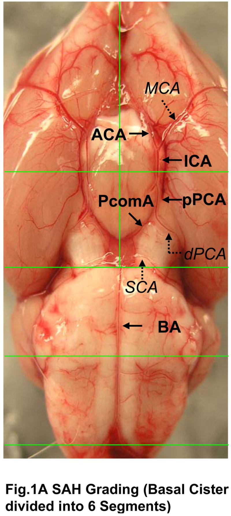
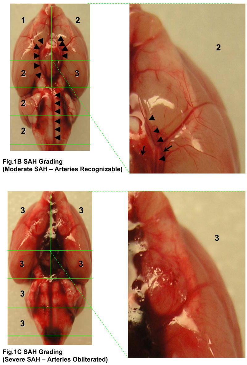
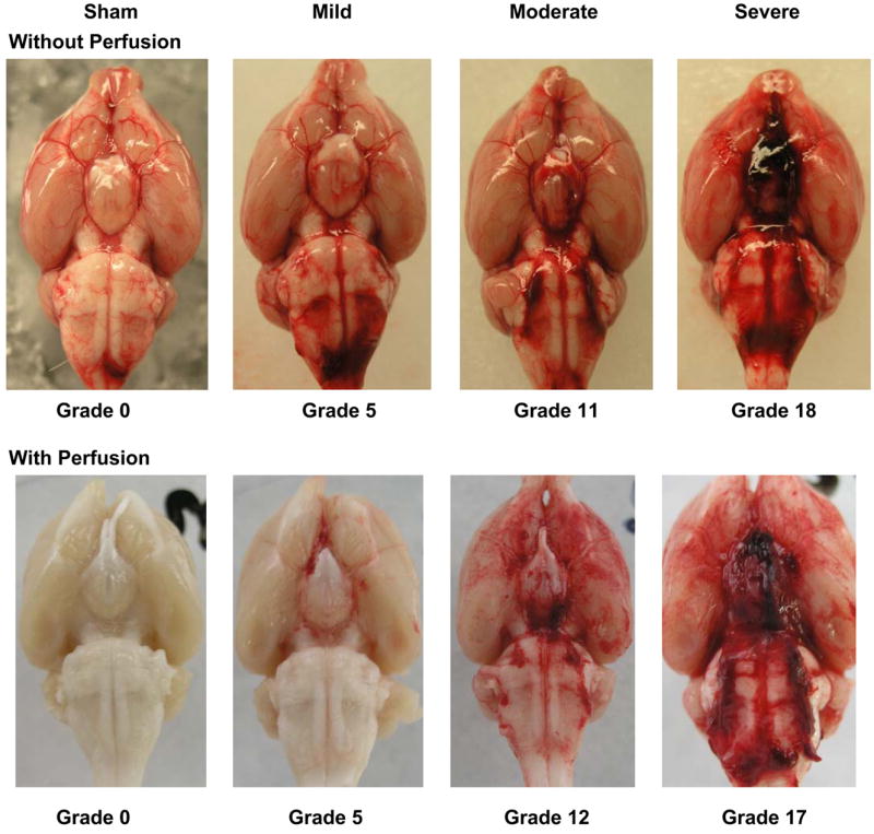
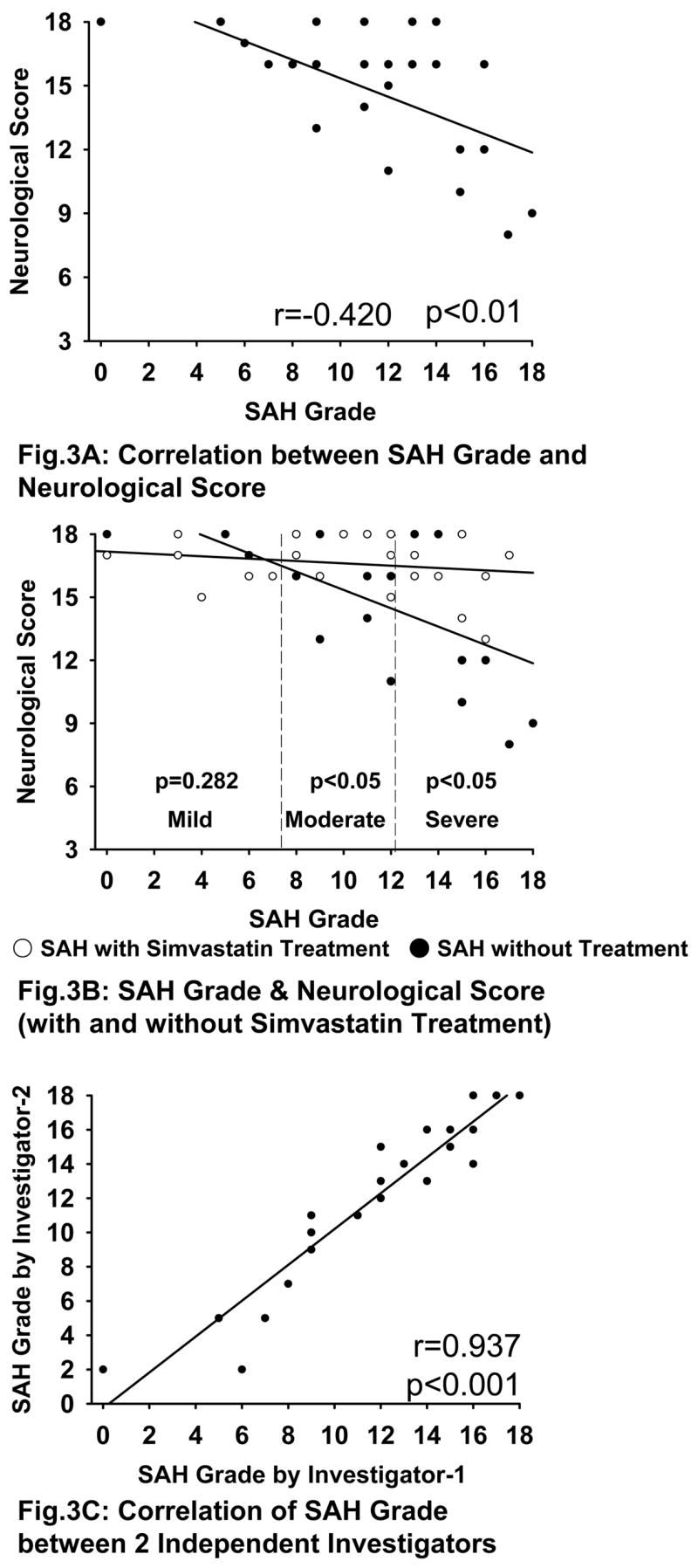
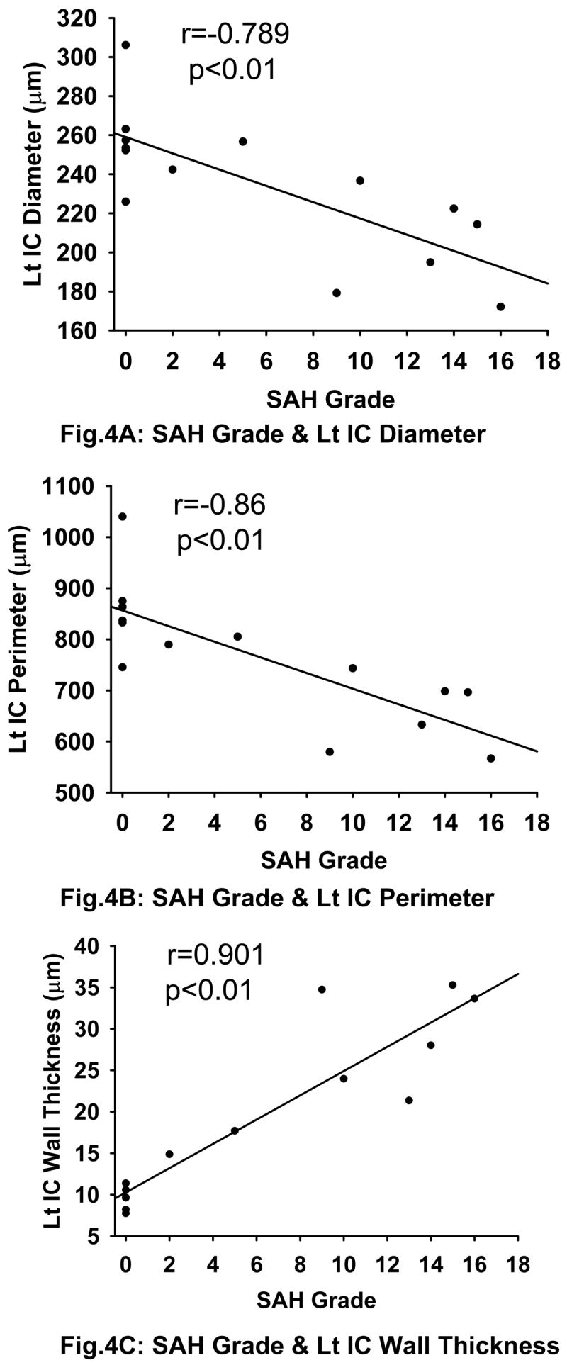
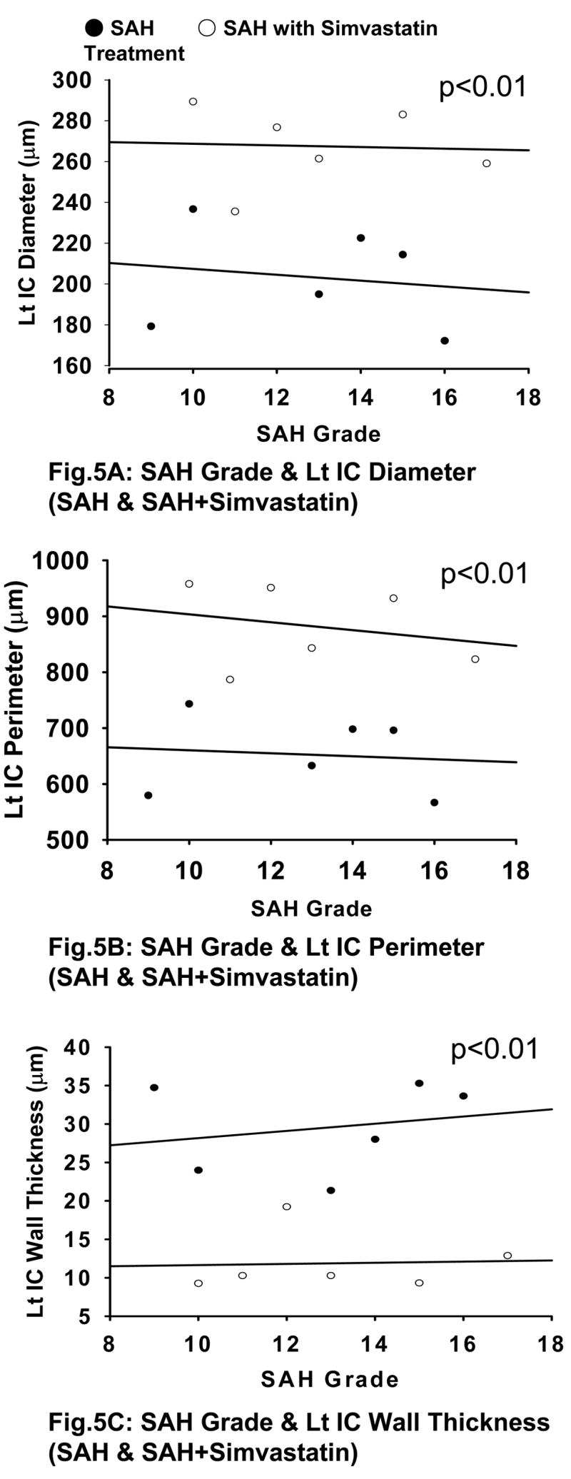
The endovascular perforation rodent model for experimental subarachnoid hemorrhage (SAH) studies is criticized for lack of control over bleeding. Presently, there is no practical grading system to categorize the severity of SAH depending on the amount of blood. We outline a simple and objective novel SAH grading system by examining the subarachnoid blood clots in the basal cisterns, and evaluate for correlation with neurological status and cerebral vasospasm. Effects of simvastatin, known to reduce vasospasm, were examined using this grading system. Seventy-seven adult male Sprague-Dawley rats were divided randomly into three groups: sham-operated (n=24), SAH (n=32), and SAH+simvastatin (n=25). High-resolution brain pictures were used to grade the severity of SAH and categorize animals into mild, moderate and severe groups. The SAH grades were compared with neurological scores and internal carotid artery parameters such as diameter, perimeter and wall thickness at 24h. Two investigators verified the grading system independently. The SAH grade showed linear correlation functionally with neurological status (r=0.42, p<0.01) and morphometrically with the degree of vasospasm (|r|>0.7, p<0.01), and also between two independent investigators (r=0.937, p<0.001). Simvastatin improved neurological score in moderate and severe (p<0.05) but not mild SAH groups (p=0.28). This grading system has the potential to be adopted for SAH experimental rodent models.


Figures

Fig 1
Fig 1
Photographs depicting SAH Grading. Fig 1A shows the division of the basal cistern into 6 segments. The subarachnoid blood clots are assessed in each of these segments to allot a score from 0–3 further explained in table 1. The arteries we identified in the basal cistern are as follows: the BA and circle of Willis composed of ACA, ICA, proximal PCA (from ICA to PcomA), and PcomA (indicated by black arrows). The MCA, distal PCA, and SCA (italicized and indicated by dotted arrows) were not used for the grading system. Fig 1B shows a representative moderate SAH. The arteries can be recognized in the magnified segment (marked by arrowheads). Arrows indicate the subarachnoid blood clots. The arteries are completely obliterated by the blood clot in Fig 1C. ACA; anterior cerebral artery, BA; basilar artery, ICA; internal carotid artery, MCA; middle cerebral artery, PCA; posterior cerebral artery, PcomA; posterior communicating artery, SCA; superior cerebellar artery.
Fig 1
Fig 1
Photographs depicting SAH Grading. Fig 1A shows the division of the basal cistern into 6 segments. The subarachnoid blood clots are assessed in each of these segments to allot a score from 0–3 further explained in table 1. The arteries we identified in the basal cistern are as follows: the BA and circle of Willis composed of ACA, ICA, proximal PCA (from ICA to PcomA), and PcomA (indicated by black arrows). The MCA, distal PCA, and SCA (italicized and indicated by dotted arrows) were not used for the grading system. Fig 1B shows a representative moderate SAH. The arteries can be recognized in the magnified segment (marked by arrowheads). Arrows indicate the subarachnoid blood clots. The arteries are completely obliterated by the blood clot in Fig 1C. ACA; anterior cerebral artery, BA; basilar artery, ICA; internal carotid artery, MCA; middle cerebral artery, PCA; posterior cerebral artery, PcomA; posterior communicating artery, SCA; superior cerebellar artery.
Fig. 2
Fig. 2
Degree of SAH Severity. Figure shows representative pictures of sham, mild, moderate and severe SAH groups with and without perfusion.
Fig. 3
Fig. 3
Correlation of SAH Grade with Neurological Score and between independent investigators. Fig 3A shows that there is a significant correlation between SAH grade and neurological score (r=0.42, p<0.01). Fig 3B shows a significant difference between SAH+vehicle and SAH+simvastatin groups in over all SAH grade (p<0.05). There is also significant difference between these groups in moderate (p<0.05) and severe (p<0.05) SAH but not mild SAH (p=0.282). Fig 3C shows a significantly strong linear correlation for SAH grading between two independent investigators (r=0.937, p<0.001).
Fig. 4
Fig. 4
Correlation between SAH Grade and Cerebral Vasospasm. There is a strong linear correlation between the SAH grade and intracranial IC diameter (Fig 4A) (r=−0.789, p<0.01), perimeter (Fig 4B) (r=−0.860, p<0.01) and wall thickness (Fig 4C) (r=0.901, p<0.01). All the three figures also include data from the sham group which is clustered at grade 0 on the x-axis as expected. Lt IC indicates the left / ipsilateral internal carotid artery.
Fig. 5
Fig. 5
SAH Grade and Cerebral Vasospasm (with and without simvastatin treatment). There is a significant difference between SAH and SAH+simvastatin groups in IC diameter (Fig 5A), IC perimeter (Fig 5B), and IC wall thickness (Fig 5C) (p<0.01). Lt IC indicates the left / ipsilateral internal carotid artery.

References

    1. Aladag MA, Turkoz Y, Ozcan C, Sahna E, Parlakpinar H, Akpolat N, et al. Caffeic acid phenethyl ester (CAPE) attenuates cerebral vasospasm after experimental subarachnoidal haemorrhage by increasing brain nitric oxide levels. Int J Dev Neurosci. 2006;24:9–14. - PubMed
    1. Bederson JB, Germano IM, Guarino L. Cortical blood flow and cerebral perfusion pressure in a new noncraniotomy model of subarachnoid hemorrhage in the rat. Stroke. 1995;26:1086–1091. - PubMed
    1. Cahill J, Calvert JW, Solaroglu I, Zhang JH. Vasospasm and p53-induced apoptosis in an experimental model of subarachnoid hemorrhage. Stroke. 2006;37:1868–1874. - PubMed
    1. Cimino M, Balduini W, Carloni S, Gelosa P, Guerrini U, Tremoli E, et al. Neuroprotective effect of simvastatin in stroke: a comparison between adult and neonatal rat models of cerebral ischemia. Neurotoxicology. 2005;26:929–933. - PubMed
    1. Garcia JH, Wagner S, Liu KF, Hu XJ. Neurological deficit and extent of neuronal necrosis attributable to middle cerebral artery occlusion in rats. Statistical validation Stroke. 1995;26:627–634. - PubMed

Publication types

MeSH terms

Substances