Review
. 2007 Aug;17(4):427-36.
doi: 10.1016/j.sbi.2007.08.016. Epub 2007 Sep 17.

Structural basis of viral invasion: lessons from paramyxovirus F

Affiliations
Review

Structural basis of viral invasion: lessons from paramyxovirus F

Robert A Lamb et al. Curr Opin Struct Biol. 2007 Aug.

Abstract

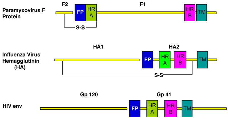
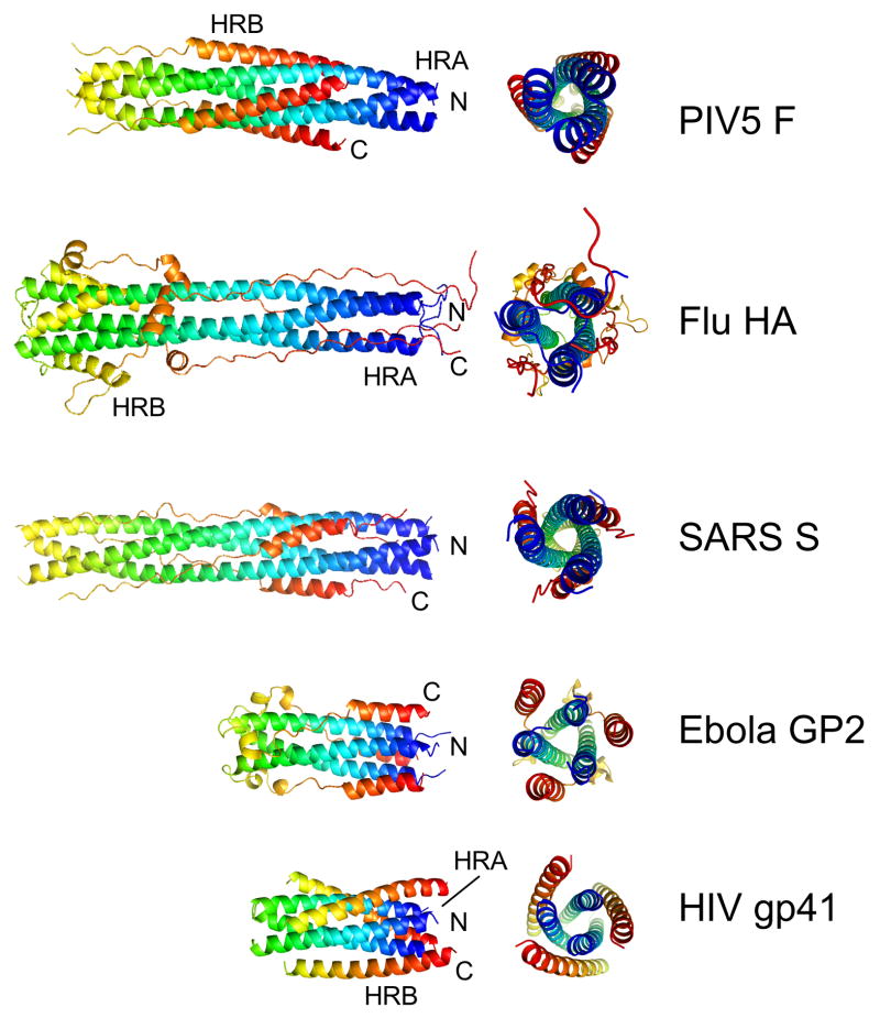
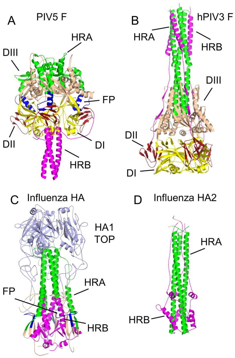
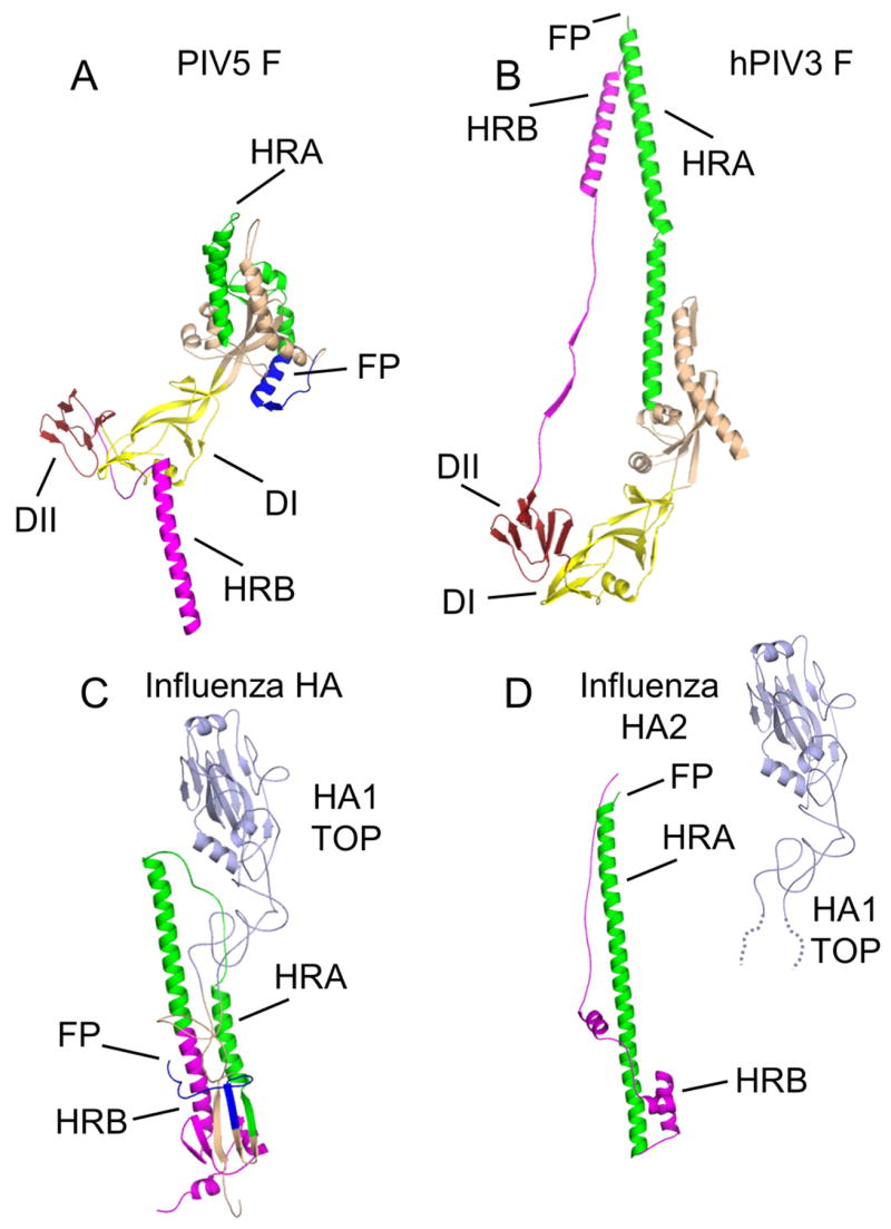
The structures of glycoproteins that mediate enveloped virus entry into cells have revealed dramatic structural changes that accompany membrane fusion and provided mechanistic insights into this process. The group of class I viral fusion proteins includes the influenza hemagglutinin, paramyxovirus F, HIV env, and other mechanistically related fusogens, but these proteins are unrelated in sequence and exhibit clearly distinct structural features. Recently determined crystal structures of the paramyxovirus F protein in two conformations, representing pre-fusion and post-fusion states, reveal a novel protein architecture that undergoes large-scale, irreversible refolding during membrane fusion, extending our understanding of this diverse group of membrane fusion machines.


Figures

Figure 1
Figure 1. Schematic of three prototypical class I viral fusion proteins
Schematics of the paramyxovirus F, influenza virus HA and HIV env glycoproteins are shown with important sequence features annotated. All three proteins are produced as intact polypeptide chains, which form trimers that are subsequently cleaved (gap) during biosynthesis. Paramyxovirus F is cleaved into two subunits, F1 and F2, HA into HA1 and HA2 and HIV env into gp120 and gp41. The site of cleavage is adjacent to a hydrophobic stretch of 20–25 amino acids known as the fusion peptide (FP; blue) followed by an N-terminal heptad repeat sequence (HRA; green). In paramyxovirus F and HIV env, a separate C-terminal heptad repeat sequence is easily identifiable (HRB; magenta). In the influenza HA sequence, we have used HRB to indicate the C-terminal portion of the primary heptad repeat that is observed to reverse its orientation during the low pH conformational change. Transmembrane anchors (TM) are also indicated and these are proximal to HRB. In paramyxovirus F, 250 amino acids separate HRA and HRB, in contrast to their much closer positioning in HA and env sequences.
Figure 2
Figure 2. Core structures of class I viral fusion proteins
Representative core structures of class I viral fusion proteins, corresponding to the HRA and HRB regions in Figure 1, are shown in their presumed postfusion conformations. The paramyxovirus F (PIV5), influenza HA2 (bacterially expressed), SARS spike glycoprotein (S), Ebola GP2 and HIV gp41 form similar hairpin-like arrangements, though with clear differences in the details of their structures. The chains are colored from blue (N-terminus) to red (C-terminus), with the blue tips of the coiled coils corresponding to the positions of the fusion peptides and the red C-termini corresponding to the positions of the TM domains. In all proteins, these membrane interacting segments are localized to the same end of the structures.
Figure 3
Figure 3. Structures of paramyxovirus F and influenza HA trimers in two conformations
(A) The prefusion conformation of the PIV5 F trimer. HRA is colored green, HRB and its N-terminal linker region are colored magenta and the fusion peptides are colored blue. Domains I (DI), II (DII) and III (DIII) are indicated. The DI domains, which remain relatively invariant during the conformational change, are shown in yellow and DII domains are in dark red. (B) The postfusion conformation of the hPIV3 F trimer. Coloring of the protein is as for the PIV5 F trimer in (A). (C) The prefusion conformation of the influenza HA trimer. The coloring of HRA, HRB (and its C-terminal linker to the TM domain), and the fusion peptides follow those used for the PIV5 trimer in (A). The region of HA1 that forms a globular receptor-binding domain (HA1 TOP), is colored light blue. (D) The postfusion conformation of the influenza HA trimer, as revealed by the crystal structure of the C-terminal HA2 subunit. Coloring of the protein is as in (C). The HA1 subunit, including HA1 TOP, is thought to dissociate from HA2, becoming linked by disordered and protease-sensitive tethers during exposure to low pH.
Figure 4
Figure 4. Structures of paramyxovirus F and influenza HA subunits in two conformations
(A) The prefusion conformation of a single PIV5 F subunit. (B) The postfusion conformation of a single hPIV3 F subunit. (C) The prefusion conformation of a single influenza HA subunit. (D) The postfusion conformation of a single subunit of the influenza HA2 protein. The flexible linkage of HA2 to HA1 TOP is indicated by dotted lines representing amino acids present in HA1, that extend from the globular HA1 TOP domain. The HA1–HA2 disulfide bond is omitted for clarity. Coloring of the subunits follows those used in Figure 3.
Figure 5
Figure 5. Electron microscopy of the PIV5 F protein and progression from the pre-fusion to the post-fusion conformation
Top Panel. (A). F-GCNt, (B) F-GCNt trypsin digested, (C) F-GCNt heated to 50°C for 30 min, (D) F-GCNt digested with trypsin and then heated to 50°C for 30 min, (E) F-GCNt heated to 50°C for 30 min and then digested with trypsin. Bottom panel: The unheated and heated proteins are depicted by the PIV5 F-GCNt (Protein data bank [PDB] 2B9B) and hPIV3 solF0 (PDB 1ZTM) structures, respectively. The HRA (red), fusion peptide (green), HRB (blue), and GCNt (magenta) regions are colored. When F-GCNt is cleaved with trypsin (left side), the protein does not refold to its post-fusion conformation, but a gain of MAb F1a reactivity indicates a subtle change. When cleaved F-GCNt is heated, the protein converts to the post-fusion golf tee-like conformation, aggregates into rosettes through its fusion peptide, and gains MAb 6–7 reactivity. If C1 peptide (HRA) is added during the heating of cleaved F-GCNt, it can bind to the protein and most likely trap the pre-hairpin intermediate. When F-GCNt is heated without cleavage (right side), the protein refolds into its uncleaved post-fusion conformation. The 47 residues for which there is no interpretable density in the hPIV3 electron density map, including the residues encoding the cleavage site and fusion peptide, have been added as dotted lines. When heated F-GCNt is cleaved, the protein aggregates into rosettes. It is anticipated that the TM domain or GCNt domain would be adjacent to the fusion peptide in rosettes, but for clarity this has been omitted.

Similar articles

Cited by

References

    1. Harrison SC. Principles of Virus Structure. In: Knipe DM, Howley PM, editors. Fields Virology. 5. Vol. 1 Wolters Kluwer: Lippincott WIlliams & Wilkins; 2007. pp. 59–98.
    1. Helenius A. Virus Entry and Uncoating. In: Knipe DM, Howley PM, editors. Fields Virology. Vol. 1 Wolters Kluwer: Lippincott WIlliams & Wilkins; 2007. pp. 99–118.
    1. Wilson IA, Skehel JJ, Wiley DC. Structure of the haemagglutinin membrane glycoprotein of influenza virus at 3 Å resolution. Nature. 1981;289:366–375. - PubMed
    1. Bullough PA, Hughson FM, Skehel JJ, Wiley DC. Structure of influenza haemagglutinin at the pH of membrane fusion. Nature. 1994;371:37–43. - PubMed
    1. Skehel JJ, Wiley DC. Receptor binding and membrane fusion in virus entry: the influenza hemagglutinin. Annu Rev Biochem. 2000;69:531–569. - PubMed

Publication types

Substances

LinkOut - more resources