

In praise of tedious anatomy
- PMID: 17870621
- PMCID: PMC1986635
- DOI: 10.1016/j.neuroimage.2006.09.055
In praise of tedious anatomy
Abstract
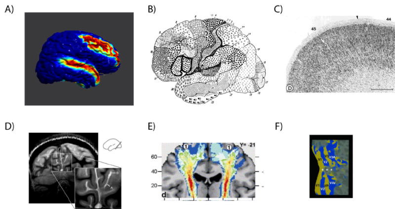
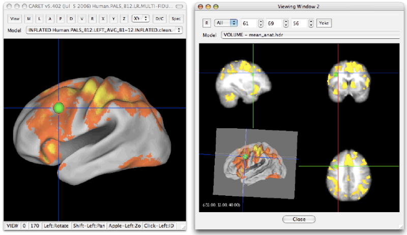
Functional neuroimaging is fundamentally a tool for mapping function to structure, and its success consequently requires neuroanatomical precision and accuracy. Here we review the various means by which functional activation can be localised to neuroanatomy and suggest that the gold standard should be localisation to the individual's or group's own anatomy through the use of neuroanatomical knowledge and atlases of neuroanatomy. While automated means of localisation may be useful, they cannot provide the necessary accuracy, given variability between individuals. We also suggest that the field of functional neuroimaging needs to converge on a common set of methods for reporting functional localisation including a common "standard" space and criteria for what constitutes sufficient evidence to report activation in terms of Brodmann's areas.
Figures
Comment in
-
Functional magnetic resonance imaging: measuring versus estimating.Neuroimage. 2007 Oct 1;37(4):1042-4; discussion 1066-8. doi: 10.1016/j.neuroimage.2007.02.038. Epub 2007 Mar 6. Neuroimage. 2007. PMID: 17428686
-
Commentary on Devlin and Poldrack.Neuroimage. 2007 Oct 1;37(4):1055-6; discussion 1066-8. doi: 10.1016/j.neuroimage.2007.02.010. Epub 2007 Jun 8. Neuroimage. 2007. PMID: 17560127 No abstract available.
-
Comment on Devlin and Poldrack.Neuroimage. 2007 Oct 1;37(4):1057-8; discussion 1066-8. doi: 10.1016/j.neuroimage.2007.02.009. Epub 2007 Aug 27. Neuroimage. 2007. PMID: 17719800 No abstract available.
-
What is where and why it is important.Neuroimage. 2007 Oct 1;37(4):1045-9; discussion 1066-8. doi: 10.1016/j.neuroimage.2007.02.018. Epub 2007 Feb 21. Neuroimage. 2007. PMID: 17720552 Free PMC article. No abstract available.
-
On navigating the human cerebral cortex: response to 'in praise of tedious anatomy'.Neuroimage. 2007 Oct 1;37(4):1050-4; discussion 1066-8. doi: 10.1016/j.neuroimage.2007.02.021. Epub 2007 Sep 4. Neuroimage. 2007. PMID: 17766148 Free PMC article.
-
Neuroanatomy: tool for functional localization, key to brain organization.Neuroimage. 2007 Oct 1;37(4):1059-60; discussion 1066-8. doi: 10.1016/j.neuroimage.2007.02.007. Epub 2007 Sep 5. Neuroimage. 2007. PMID: 17822924
-
Cytoarchitecture of the cerebral cortex--more than localization.Neuroimage. 2007 Oct 1;37(4):1061-5; discussion 1066-8. doi: 10.1016/j.neuroimage.2007.02.037. Epub 2007 Mar 3. Neuroimage. 2007. PMID: 17870622
References
-
- Amunts K, Malikovic A, Mohlberg H, Schormann T, Zilles K. Brodmann’s areas 17 and 18 brought into stereotaxic space-where and how variable? Neuroimage. 2000;11:66–84. - PubMed
-
- Amunts K, Schleicher A, Burgel U, Mohlberg H, Uylings HB, et al. Broca’s region revisited: Cytoarchitecture and intersubject variability. J Comp Neurol. 1999;412:319–341. - PubMed
-
- Anwander A, Tittgemeyer M, von Cramon DY, Friederici AD, Knosche TR. Connectivity-based parcellation of broca’s area. Cereb Cortex 2006 - PubMed
-
- Barbier EL, Marrett S, Danek A, Vortmeyer A, van Gelderen P, et al. Imaging cortical anatomy by high-resolution mr at 3.0t: Detection of the stripe of gennari in visual area 17. Magn Reson Med. 2002;48:735–738. - PubMed
-
- Behrens TE, Johansen-Berg H, Woolrich MW, Smith SM, Wheeler-Kingshott CA, et al. Non-invasive mapping of connections between human thalamus and cortex using diffusion imaging. Nat Neurosci. 2003;6:750–757. - PubMed
Publication types
MeSH terms
Grants and funding
LinkOut - more resources
Full Text Sources
Medical
