

Arrhythmogenic mechanisms in a mouse model of catecholaminergic polymorphic ventricular tachycardia
- PMID: 17872467
- PMCID: PMC2515360
- DOI: 10.1161/CIRCRESAHA.107.148064
Arrhythmogenic mechanisms in a mouse model of catecholaminergic polymorphic ventricular tachycardia
Abstract
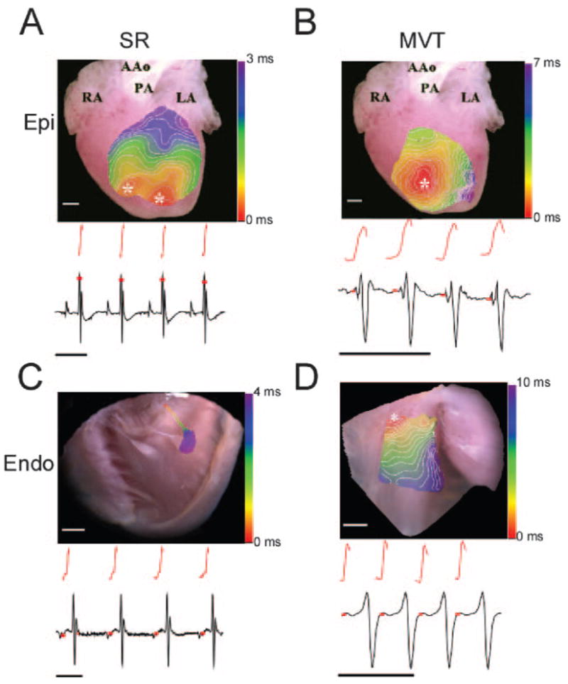
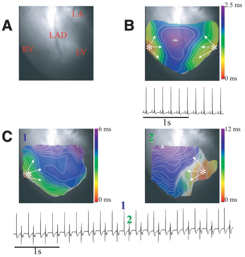
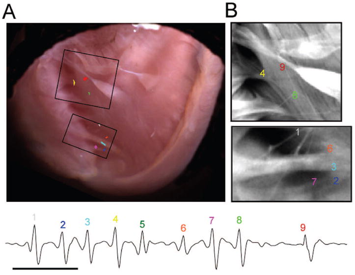
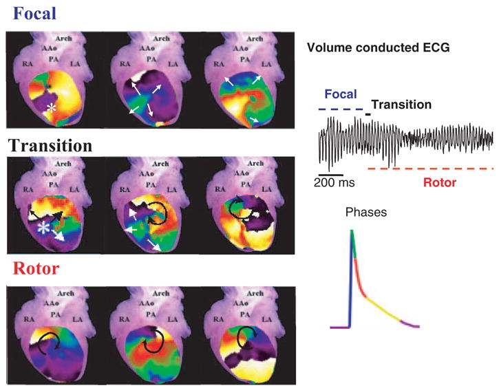
Catecholaminergic polymorphic ventricular tachycardia (VT) is a lethal familial disease characterized by bidirectional VT, polymorphic VT, and ventricular fibrillation. Catecholaminergic polymorphic VT is caused by enhanced Ca2+ release through defective ryanodine receptor (RyR2) channels. We used epicardial and endocardial optical mapping, chemical subendocardial ablation with Lugol's solution, and patch clamping in a knockin (RyR2/RyR2(R4496C)) mouse model to investigate the arrhythmogenic mechanisms in catecholaminergic polymorphic VT. In isolated hearts, spontaneous ventricular arrhythmias occurred in 54% of 13 RyR2/RyR2(R4496C) and in 9% of 11 wild-type (P=0.03) littermates perfused with Ca2+and isoproterenol; 66% of 12 RyR2/RyR2(R4496C) and 20% of 10 wild-type hearts perfused with caffeine and epinephrine showed arrhythmias (P=0.04). Epicardial mapping showed that monomorphic VT, bidirectional VT, and polymorphic VT manifested as concentric epicardial breakthrough patterns, suggesting a focal origin in the His-Purkinje networks of either or both ventricles. Monomorphic VT was clearly unifocal, whereas bidirectional VT was bifocal. Polymorphic VT was initially multifocal but eventually became reentrant and degenerated into ventricular fibrillation. Endocardial mapping confirmed the Purkinje fiber origin of the focal arrhythmias. Chemical ablation of the right ventricular endocardial cavity with Lugol's solution induced complete right bundle branch block and converted the bidirectional VT into monomorphic VT in 4 anesthetized RyR2/RyR2(R4496C) mice. Under current clamp, single Purkinje cells from RyR2/RyR2(R4496C) mouse hearts generated delayed afterdepolarization-induced triggered activity at lower frequencies and level of adrenergic stimulation than wild-type. Overall, the data demonstrate that the His-Purkinje system is an important source of focal arrhythmias in catecholaminergic polymorphic VT.
Figures


Comment in
-
The perfect storm: defective calcium cycling in insulated fibers with reduced repolarization reserve.Circ Res. 2007 Nov 9;101(10):968-70. doi: 10.1161/CIRCRESAHA.107.164426. Circ Res. 2007. PMID: 17991890 No abstract available.
References
-
- Leenhardt A, Lucet V, Denjoy I, Grau F, Ngoc DD, Coumel P. Catecholaminergic polymorphic ventricular tachycardia in children. A 7-year follow-up of 21 patients. Circulation. 1995;91:1512–1519. - PubMed
-
- Priori SG, Napolitano C, Tiso N, Memmi M, Vignati G, Bloise R, Sorrentino V, Danieli GA. Mutations in the cardiac ryanodine receptor gene (hRyR2) underlie catecholaminergic polymorphic ventricular tachycardia. Circulation. 2001;103:196–200. - PubMed
-
- Priori SG, Napolitano C, Memmi M, Colombi B, Drago F, Gasparini M, DeSimone L, Coltorti F, Bloise R, Keegan R, Cruz Filho FE, Vignati G, Benatar A, DeLogu A. Clinical and molecular characterization of patients with catecholaminergic polymorphic ventricular tachycardia. Circulation. 2002;106:69–74. - PubMed
-
- Cerrone M, Colombi B, Santoro M, di Barletta MR, Scelsi M, Villani L, Napolitano C, Priori SG. Bidirectional ventricular tachycardia and fibrillation elicited in a knockin mouse model carrier of a mutation in the cardiac ryanodine receptor. Circ Res. 2005;96:e77–e82. - PubMed
-
- Liu N, Colombi B, Memmi M, Zissimopoulos S, Rizzi N, Negri S, Imbriani M, Napolitano C, Lai FA, Priori SG. Arrhythmogenesis in catecholaminergic polymorphic ventricular tachycardia. Insights from a RyR2 R4496C knockin mouse model. Circ Res. 2006;99:292–298. - PubMed
Publication types
MeSH terms
Substances
Grants and funding
LinkOut - more resources
Full Text Sources
Other Literature Sources
Molecular Biology Databases
Miscellaneous
