Comparative Study
. 2007 Dec;72(6):1508-21.
doi: 10.1124/mol.107.038984. Epub 2007 Sep 14.

Resveratrol (trans-3,5,4'-trihydroxystilbene) ameliorates experimental allergic encephalomyelitis, primarily via induction of apoptosis in T cells involving activation of aryl hydrocarbon receptor and estrogen receptor

Affiliations
Comparative Study

Resveratrol (trans-3,5,4'-trihydroxystilbene) ameliorates experimental allergic encephalomyelitis, primarily via induction of apoptosis in T cells involving activation of aryl hydrocarbon receptor and estrogen receptor

Narendra P Singh et al. Mol Pharmacol. 2007 Dec.

Abstract

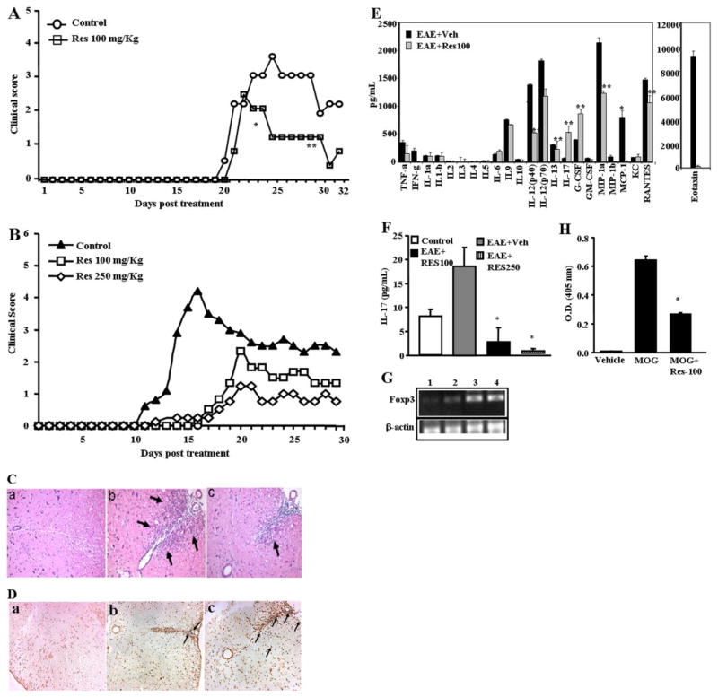
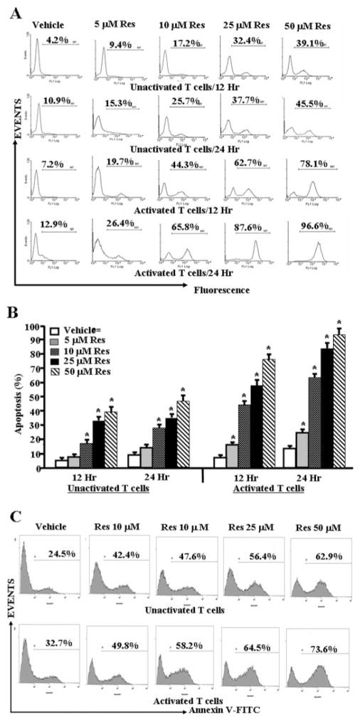
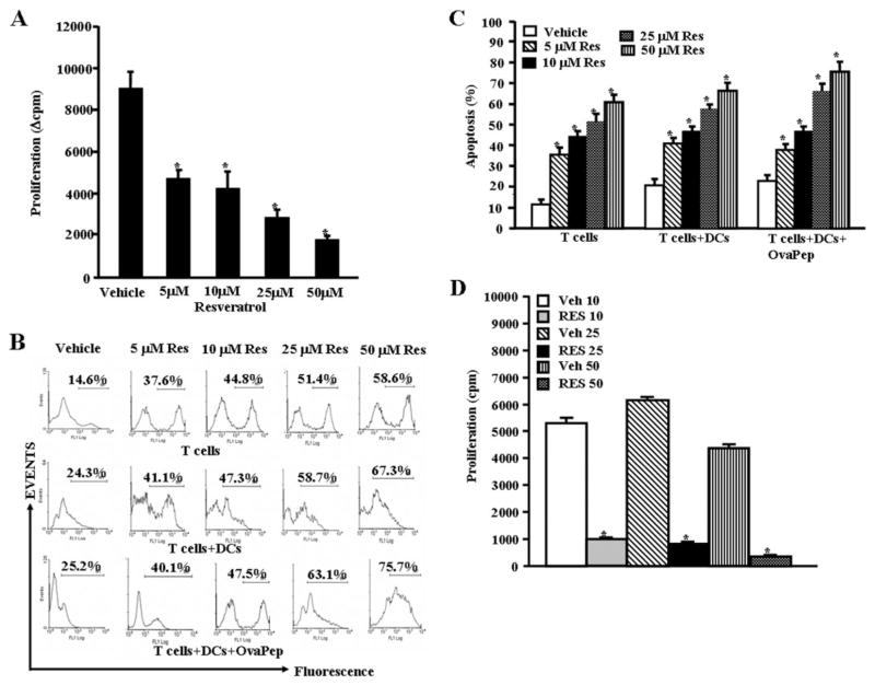
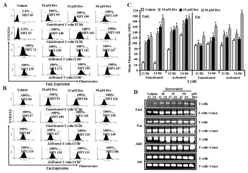
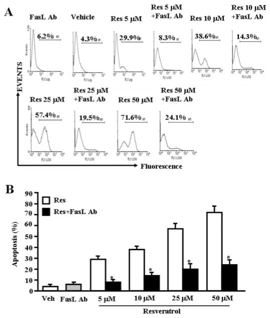
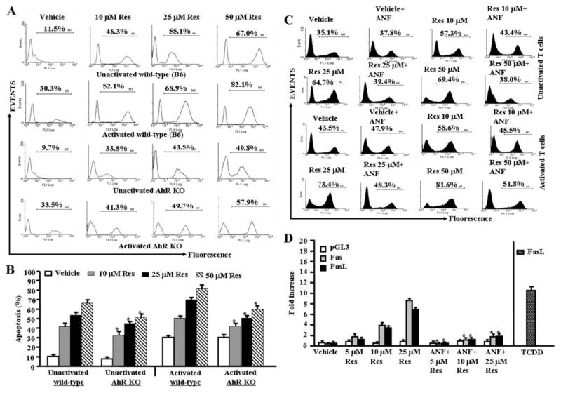
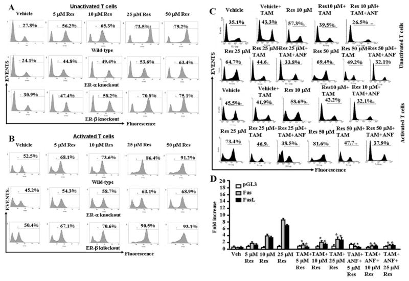
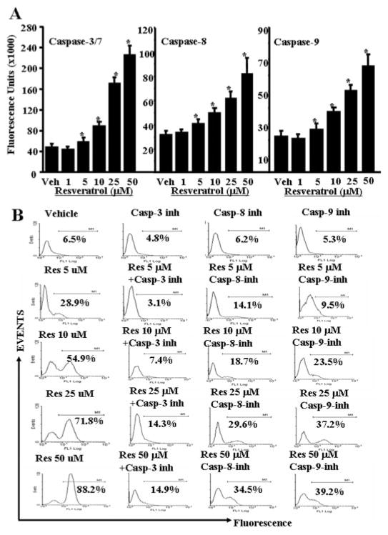
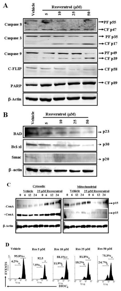
Resveratrol (trans-3,5,4'-trihydroxystilbene), a polyphenolic compound found in plant products, including red grapes, exhibits anticancer, antioxidant, and anti-inflammatory properties. Using an animal model of multiple sclerosis (MS), we investigated the use of resveratrol for the treatment of autoimmune diseases. We observed that resveratrol treatment decreased the clinical symptoms and inflammatory responses in experimental allergic encephalomyelitis (EAE)-induced mice. Furthermore, we observed significant apoptosis in inflammatory cells in spinal cord of EAE-induced mice treated with resveratrol compared with the control mice. Resveratrol administration also led to significant down-regulation of certain cytokines and chemokines in EAE-induced mice including tumor necrosis factor-alpha, interferon-gamma, interleukin (IL)-2, IL-9, IL-12, IL-17, macrophage inflammatory protein-1alpha (MIP-1alpha), monocyte chemoattractant protein-1 (MCP-1), regulated on activation normal T-cell expressed and secreted (RANTES), and Eotaxin. In vitro studies on the mechanism of action revealed that resveratrol triggered high levels of apoptosis in activated T cells and to a lesser extent in unactivated T cells. Moreover, resveratrol-induced apoptosis was mediated through activation of aryl hydrocarbon receptor (AhR) and estrogen receptor (ER) and correlated with up-regulation of AhR, Fas, and FasL expression. In addition, resveratrol-induced apoptosis in primary T cells correlated with cleavage of caspase-8, caspase-9, caspase-3, poly(ADP-ribose) polymerase, and release of cytochrome c. Data from the present study demonstrate, for the first time, the ability of resveratrol to trigger apoptosis in activated T cells and its potential use in the treatment of inflammatory and autoimmune diseases including, MS.


Figures

Fig. 1
Fig. 1
Resveratrol treatment diminishes clinical symptoms of EAE. A and B, C57BL/6 mice were immunized with 20 μg (A) or 150 μg (B) of MOG35-55 peptide emulsified in complete Freund’s adjuvant and 400 ng of pertussis toxin. These immunized mice received 100 mg/kg (A and B) or 250 mg/kg (B) of resveratrol daily from day 2 until the termination of the experiment. C, images from immunohistopathological examination of spinal cord of EAE-induced mice. Spinal cords were collected from: a, normal mice; b, EAE-induced vehicle-treated mice; c, EAE-induced resveratrol-treated mice. Arrows indicate infiltration of polymorphonuclear cells. D, in situ TUNEL assays for apoptosis in spinal cord of EAE-induced mice. Spinal cords from normal mice (a), EAE-induced vehicle-treated mice (b), EAE-induced resveratrol-treated mice (c) were collected and in situ apoptosis was determined using DeadEnd colometric TUNEL system. E, expression of various cytokines and chemokines in serum of EAE-induced mice on day 25 after immunization that were treated with vehicle or resveratrol. F, expression of cytokine IL-17 in serum of EAE-induced mice on day 9. G, expression of Foxp3 in splenocytes harvested from EAE induced mice (C57BL/6) treated with vehicle or resveratrol on day 25 after immunization. Lane 1, normal spleen; lane 2, MOG + Vehicle; lane 3, MOG + resveratrol (100 mg/kg), and lane 4: MOG + resveratrol (500 mg/kg). β-Actin was used as a positive control. H, presence of anti-MOG antibody in the sera of EAE induced mice (C57BL/6) treated with vehicle or resveratrol on day 25 after immunization. Data represent mean ± S.E.M. of six animals and asterisks (*) represent significant differences between resveratrol-treated groups compared with vehicle controls.
Fig. 2
Fig. 2
Resveratrol triggers apoptosis in primary T cells. Purified primary T cells from C57BL/6 mice were activated or not with ConA for 24 h followed by treatment with resveratrol or vehicle for 12 to 24 h (A–C). T cells were analyzed for apoptosis by TUNEL assays (A and B) and Annexin V/PI (C). A demonstrates representative experiment for apoptosis and B shows mean ± S.E.M. of four independent experiments. C demonstrates representative experiment on apoptosis determined using Annexin V/PI. Cells stained for Annexin but negative for PI alone have been depicted. We observed 5 to 6% dead (PI-positive) cells (data not shown). Asterisks (*) indicate statistically significant difference between resveratrol-treated groups compared with vehicle controls.
Fig. 3
Fig. 3
Resveratrol causes apoptosis in antigen-specific activated T cells in vitro. Purified primary T cells from OT.II.2a mice were activated with ovalbumin-specific peptide (ISQAVHAAHAEINEAGR) by coculturing T cells and peptide-pulsed mature DCs for 2 days and then treated with various doses of resveratrol (5–50 μM; A–C). Proliferation of T cells was determined by [3H]thymidine incorporation (A) 24 h after resveratrol or vehicle exposure and apoptosis as determined by TUNEL assays (B and C). A represents mean ± S.E.M. of triplicate cultures and data were expressed as change in counts per minute. Asterisks (*) represent significant difference in proliferation of antigen-specific (ova-peptides) activated OT.II.2a T cells treated with resveratrol compared with vehicle controls. B is a representative of three independent TUNEL assays, and C represents mean ± S.E.M. of three independent TUNEL experiments and asterisks (*) represent significant difference in apoptosis between resveratrol-treated groups compared with vehicle controls. D, popliteal draining lymph nodes were harvested from mice immunized with MOG (150 μg/mice) on day 7 and cultured in the presence of MOG (10 μg/ml) and vehicle or resveratrol (10–50 μM) for 3 days. Proliferation of lymph node cells was determined by [3H]thymidine incorporation (D). D represents mean ± S.E.M. of triplicate cultures. Asterisks (*) represent significant difference in proliferation of MOG-specific activated lymph node cells treated with resveratrol compared with vehicle controls.
Fig. 4
Fig. 4
Role of Fas and FasL in resveratrol-mediated apoptosis in T cells. Expression of Fas and FasL in unactivated or ConA-activated T cells was determined by staining the cells with anti-mouse Fas-PE and anti-mouse FasL-PE antibodies and analyzed by Flow cytometry and RT-PCR. A, expression of FasL in unactivated or ConA-activated T cells 12 and 24 h after resveratrol or vehicle treatment (flow cytometry). B, expression of Fas in unactivated or ConA-activated T cells 12 and 24 h after resveratrol or vehicle treatment. C, mean fluorescent intensity of FasL and Fas expression after resveratrol or vehicle treatment. C represents mean ± S.E.M. of three independent experiments and asterisks (*) represent significant difference between resveratrol-treated groups compared with vehicle controls. D, expression of AhR, Fas, and FasL in unactivated T cells and activated T cells (T cells + ConA) using RT-PCR. 18S, a housekeeping gene, was used as a positive control.
Fig. 5
Fig. 5
FasL plays critical role in induction of death-receptor pathway of apoptosis by resveratrol. T cells purified from C57BL/6 (wild-type) mice were activated with ConA and cultured in the presence of vehicle or resveratrol (5–50 μM) and incubated in the absence or presence of mouse-specific anti-FasL Ab. Apoptosis in T cells was determined by TUNEL assays. The data presented in A are representative of three independent experiments. B represents mean of three independent experiments, and asterisks (*) represent significant reduction in resveratrol-induced apoptosis of T cells cultured in the presence of FasL Ab compared with the controls.
Fig. 6
Fig. 6
Role of AhR in resveratrol-induced apoptosis in T cells. A, unactivated or ConA-activated purified T cells from AhR wild-type or AhR KO mice were treated with vehicle or resveratrol, and apoptosis was determined by performing TUNEL assays as described in Fig. 2. B, mean ± S.E.M. of three independent experiments; asterisks (*) represent significant difference of resveratrol-induced apoptosis in AhR KO T cells compared with wild-type T cells. C, unactivated or ConA-activated T cells were cultured with resveratrol (10–50 μM) or vehicle (DMSO) for 24 h in the absence or presence of ANF in the culture and TUNEL assays were performed. The data presented are representative for 3 independent assays. D, ConA-activated EL4 cells were transfected with pGL-3-Fas or pGL-3-FasL promoter, cultured in the presence of resveratrol (5–25 μM) or vehicle for 24 h in the absence or presence of ANF in the culture, and luciferase assays were performed. Data represent mean of three independent experiments, and asterisks (*) indicate significant down-regulation of luciferase expression in the presence of ANF compared with cultures that did not receive ANF. Expression of luciferase in the presence of FasL promoter and treated with TCDD represents positive control.
Fig. 7
Fig. 7
Role of ER in resveratrol-induced apoptosis in T cells. A, unactivated T cells from wild-type (C57BL/6), ER-α knockout, ER-β knockout mice were cultured in the presence of various concentration (5–50 μM) of resveratrol (Res) or vehicle (DMSO) for 24 h, and TUNEL assays were performed 24 h after treatment. The data presented are representative of three independent assays. B, ConA-activated T cells from wild-type (C57BL/6), ER-α knockout, and ER-β knockout mice were cultured in the presence of various concentration (5–50 μM) of resveratrol (Res) or vehicle (DMSO) for 24 h, and TUNEL assays were performed 24 h after treatment. The data presented are representative of three independent assays. C, unactivated or ConA-activated T cells were cultured with resveratrol (10–50 μM) or vehicle (DMSO) for 24 h in the absence or presence of TAM alone or in combination with ANF (TAM+ANF) in the culture and TUNEL assays were performed. The data presented are representative for three independent assays. D, ConA-activated EL4 cells were transfected with pGL-3-Fas or pGL-3-FasL promoter, cultured in the presence of resveratrol (5–25 μM) or vehicle for 24 h in the absence or presence of TAM alone or in combination of ANF (TAM+ANF) in the culture, and luciferase assays were performed. D, mean ± S.E.M. of three independent experiments; asterisks (*) represent significant down-regulation of luciferase expression compared with controls.
Fig. 8
Fig. 8
Resveratrol activates and cleaves various caspases to cause apoptosis in primary T cells. Enzymatic activities of caspase-3/7, -8, and -9 were determined in ConA-activated primary T cells 24 h after resveratrol treatment. A, caspase-3/7, -8, and -9 activities, and the data represent mean of three independent experiments. Vertical bars represent S.E. of three experiments. Asterisks (*) represent statistically significant (p < 0.02) increase in enzymatic activities of caspase-3, -8, and -9 in resveratrol-treated groups compared with vehicle-treated T cells. B, ConA-activated T cells were cultured with vehicle or resveratrol in the presence or absence of inhibitors of caspase-3, -8, and -9. The data presented are representative of three independent experiments.
Fig. 9
Fig. 9
Resveratrol activates members of death-receptor and mitochondrial pathways. Cell lysates prepared from resveratrol-treated (24 h) unactivated primary T cells (A and B) and unactivated (−ConA) or activated primary T cells (+ConA) (C) were resolved by SDS-polyacrylamide gel electrophoresis and probed with primary Abs against various appropriate molecules. A and B, cell lysates prepared from unactivated and resveratrol-treated T cells were probed with caspase-8, caspase-3, caspase-9, caspase-2, c-FLIP, PARP (A), Bad, Bcl-xl, and Smac (B) antibodies. C, cell lysates prepared from unactivated and ConA-activated and resveratrol-treated T cells were probed with cytochrome c antibodies. β-Actin was used as a control. D, effect of resveratrol on Δψm in primary T cells. Unactivated primary T cells were treated with vehicle (DMSO) or resveratrol (5–50 μM) and stained with DIOC6 to evaluate Δψm. Results are representative of at least three independent experiments.

References

    1. Atten MJ, Godoy-Romero E, Attar BM, Milson T, Zopel M, Holian O. Resveratrol regulates cellular PKC alpha and delta to inhibit growth and induce apoptosis in gastric cancer cells. Invest New Drugs. 2005;23:111–119. - PubMed
    1. Baur JA, Pearson KJ, Price NL, Jamieson HA, Lerin C, Kalra A, Prabhu VV, Allard JS, Lopez-Lluch G, Lewis K, et al. Resveratrol improves health and survival of mice on a high-calorie diet. Nature. 2006;444:337–342. - PMC - PubMed
    1. Baur JA, Sinclair DA. Therapeutic potential of resveratrol: the in vivo evidence. Nat Rev Drug Discov. 2006;5:493–506. - PubMed
    1. Bernhard D, Tinhofer I, Tonko M, Hubl H, Ausserlechner MJ, Greil R, Kofler R, Csordas A. Resveratrol causes arrest in the S-phase prior to Fas-independent apoptosis in CEM-C7H2 acute leukemia cells. Cell Death Differ. 2000;7:834–842. - PubMed
    1. Bhat KP, Lantvit D, Christov K, Mehta RG, Moon RC, Pezzuto JM. Estrogenic and antiestrogenic properties of resveratrol in mammary tumor models. Cancer Res. 2001;61:7456–7463. - PubMed

Publication types

MeSH terms