. 2007 Nov;171(5):1713-23.
doi: 10.2353/ajpath.2007.070021. Epub 2007 Sep 14.

Microparticles of human atherosclerotic plaques enhance the shedding of the tumor necrosis factor-alpha converting enzyme/ADAM17 substrates, tumor necrosis factor and tumor necrosis factor receptor-1

Affiliations

Microparticles of human atherosclerotic plaques enhance the shedding of the tumor necrosis factor-alpha converting enzyme/ADAM17 substrates, tumor necrosis factor and tumor necrosis factor receptor-1

Matthias Canault et al. Am J Pathol. 2007 Nov.

Abstract

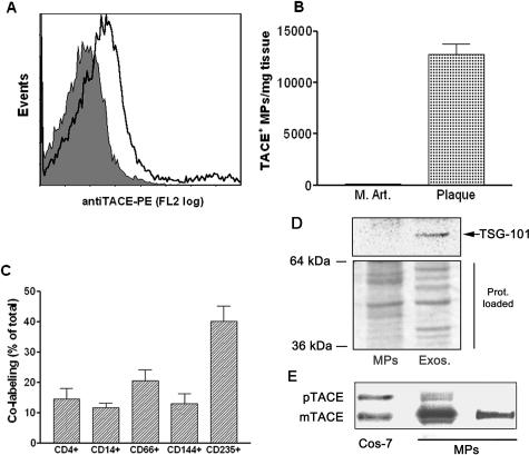
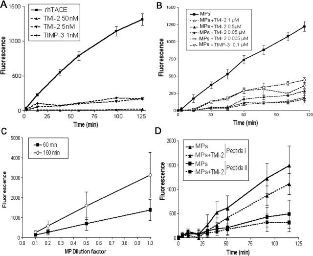
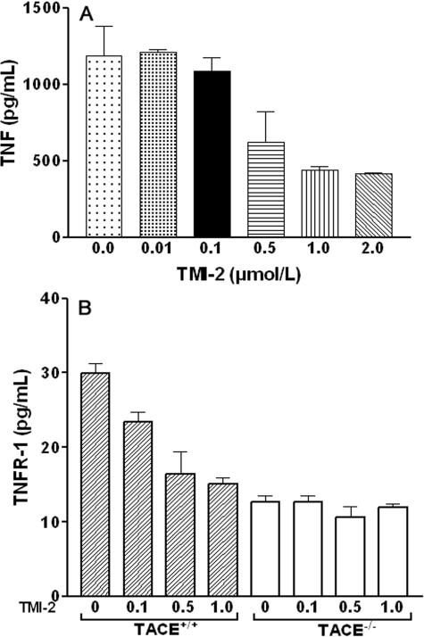
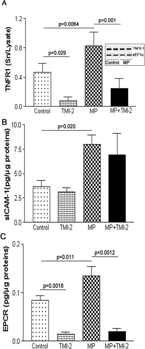
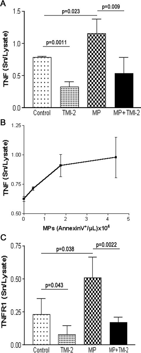
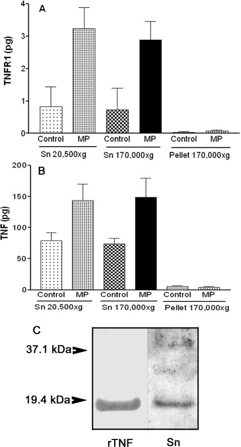
Human atherosclerotic plaques express the metalloprotease tumor necrosis factor (TNF)-alpha converting enzyme (TACE/ADAM-17), which cleaves several transmembrane proteins including TNF and its receptors (TNFR-1 and TNFR-2). Plaques also harbor submicron vesicles (microparticles, MPs) released from plasma membranes after cell activation or apoptosis. We sought to examine whether TACE/ADAM17 is present on human plaque MPs and whether these MPs would affect TNF and TNFR-1 cellular shedding. Flow cytometry analysis detected 12,867 +/- 2007 TACE/ADAM17(+) MPs/mg of plaques isolated from 25 patients undergoing endarterectomy but none in healthy human internal mammary arteries. Plaque MPs harbored mainly mature active TACE/ADAM17 and dose dependently cleaved a pro-TNF mimetic peptide, whereas a preferential TACE/ADAM17 inhibitor (TMI-2) and recombinant TIMP-3 prevented this cleavage. Plaque MPs increased TNF shedding from the human cell line ECV-304 overexpressing TNF (ECV-304(TNF)), as well as TNFR-1 shedding from activated human umbilical vein endothelial cells or ECV-304(TNF) cells, without affecting TNF or TNFR-1 synthesis. MPs also activated the shedding of the endothelial protein C receptor from human umbilical vein endothelial cells. All these effects were inhibited by TMI-2. The present study shows that human plaque MPs carry catalytically active TACE/ADAM17 and significantly enhance the cell surface processing of the TACE/ADAM17 substrates TNF, TNFR-1, and endothelial protein C receptor, suggesting that TACE/ADAM17(+) MPs could regulate the inflammatory balance in the culprit lesion.


Figures

Figure 1
Figure 1
MPs isolated from atherosclerotic human plaque, which contain the mature form of TACE/ADAM17, are of diverse cellular origin and do not contain exosomes: Analysis of TACE/ADAM17 on MPs isolated from human atherosclerotic plaque and human internal mammary arteries. A: Expression of TACE/ADAM17 on MPs from plaque homogenates. This graph is representative of the different plaque preparations. The shaded peak corresponds to negative isotype control. B: Levels of TACE/ADAM17+ MPs in human internal mammary arteries (M.Art., n = 3) and atherosclerotic plaque (plaque, n = 25); values are mean ± SEM. C: Co-labeling of TACE/ADAM17+ MPs with various cellular markers (from left to right: lymphocytes, monocytes, granulocytes, endothelial cells, and erythrocytes) (n = 12). Results are expressed as percentage of total TACE/ADAM17+ MPs (mean ± SEM). D: Immunoblotting of the exosomal marker TSG-101 in the post 20,500 × g pellet (left) and corresponding supernatant further centrifuged at 170,000 × g (right). Because protein profiles were different in both fractions (see Ponceau red staining), two times more MP materials (20 μg) than exosomal-like material were loaded. Representative of three different samples analyzed. E: Immunoblotting of TACE/ADAM17 from two different MP preparations containing 2.8 × 10⁶ and 0.1 × 10⁶ Annexin V+ MPs/μl on the middle and right lanes, respectively. The TACE/ADAM17 of MPs in the middle lane is to illustrate that only a highly MP-enriched plaque allows the detection of TACE proform. The right lane is representative of the five MP preparations tested. mTACE and pTACE indicate the positions of the mature and proform of TACE/ADAM17, respectively, validated by the migration of these forms present in COS-7 cells.
Figure 2
Figure 2
MPs isolated from human atherosclerotic plaque are active on fluorogenic peptides that are substrates of TACE/ADAM17 or MMPs. Proteolytic activity of recombinant human TACE/ADAM17 and MPs were measured on various fluorogenic substrates. Details of assay conditions are indicated in Materials and Methods. A: Time-dependent cleavage by recombinant TACE/ADAM17 of the fluorogenic peptide III, mimetic of the cleavage zone of pro-TNF (10 ng) in the presence or not of TMI-2 at 5 and 50 nmol/L or TIMP-3 (100 nmol/L), mean ± SD of two separate measurements. B: Time-dependent cleavage by MPs (10 μl) of the fluorogenic peptide III in the presence of various concentrations of TMI-2, and TIMP-3. For the sake of clarity, data are presented only as the dose of 100 nmol/L TIMP-3 because 200 nmol/L gave similar inhibition, and SEM in place of SD to avoid overlapping of error bars. n = 4. C: Dose-dependent effect of MPs [expressed as fold of the maximal amount used (10 μl)] on the cleavage of the fluorogenic peptide III. For clarity of the graph, only two time points are presented. Values are mean ± SD of two separate MP preparations. D: Time-dependent cleavage by MPs of fluorogenic peptide I and peptide II in the presence or not of TMI-2 (1 μmol/L). Values are mean ± SD of four separate MP preparations identical to those used in B.
Figure 3
Figure 3
Effects of TMI-2 on TNF and TNFR-1 release in cell-based conditions. A: ECV-304TNF cells were preincubated for 10 minutes with TMI-2 at concentrations ranging from 0 to 2 μmol/L and then stimulated with PMA (200 nmol/L) for 1 hour. Culture medium was collected for TNF assay. Values are mean ± SD of two separate experiments each performed in duplicate. B: Murine TACE/ADAM17+/+ and TACE/ADAM17ΔZn/ΔZn monocytic cells were preincubated with TMI-2 at concentrations ranging from 0 to 1.0 μmol/L and then stimulated with PMA (200 nmol/L) for 1 hour. Culture medium was collected for TNFR-1 assay. Values are mean ± SD of two separate experiments each performed in triplicate.
Figure 4
Figure 4
MPs isolated from human atherosclerotic plaque activate the release of TNFR-1, ICAM-1, and EPCR from HUVECs. HUVECs were incubated with MPs as described in Materials and Methods. Release in the culture medium of TNFR-1 (A) (values are mean ± SD, n = 12, each in duplicate), ICAM-1 (B) (values are mean ± SD, n = 3, each in duplicate), and EPCR (C) (values are mean ± SD, n = 8, each in duplicate). Significance of MP and TMI-2 effects was calculated by t-test (Mann-Whitney U-test).
Figure 5
Figure 5
MPs isolated from human atherosclerotic plaque activate the release of TNF and TNFR-1 from the human cell line ECV-304TNF. ECV-304TNF cells were incubated with MPs as described in Materials and Methods. A: Release of TNF. Values are mean ± SD (n = 6, each performed in duplicate). Significance of MP and TMI-2 effects was calculated by t-test (Mann-Whitney U-test). B: Dose-dependent effect of MPs (expressed as Annexin V+/μl) on TNF release. C: Release of TNFR-1 measured on the same samples as in A.
Figure 6
Figure 6
Plaque MPs do not induce the release of exosome-associated full-length TNFR-1 and TNF from endothelial cells. HUVECs (A) or ECV-304TNF cells (B) were incubated with or without plaque MPs as indicated in Figures 4 and 5, respectively. The culture medium was centrifuged at 20,500 × g (45 minutes at 4°C) to pellet MPs, and half of the resulting supernatant (Sn 20,500 × g) was further centrifuged at 170,000 × g (16 hours at 4°C) to pellet exosomes and the other half left for the same time at 4°C. The two supernatants (Sn 20,500 × g; Sn 170,000 × g) and the 170,000 × g pellet were assayed for TNFR-1 (expressed as total amount, pg). For both cellular types, at least 98% of TNFR-1 or TNF was recovered in the 170,000 × g supernatant (Sn). Values are mean ± SD; n = 3. C: The culture medium of ECV-304TNF cells exposed to plaque MPs (Sn) contain only the cleaved form of TNF compared with 10 μg of recombinant human TNF (rTNF) on the left lane. Representative of two different samples analyzed.

References

    1. Hansson GK. Inflammation, atherosclerosis, and coronary artery disease. N Engl J Med. 2005;352:1685–1695. - PubMed
    1. Kockx MM, De Meyer GRY, Muhring J, Jacob W, Bult H, Herman AG. Apoptosis and related proteins in different stages of human atherosclerotic plaques. Circulation. 1998;97:2307–2315. - PubMed
    1. Mallat Z, Tedgui A. Current perspective on the role of apoptosis in atherothrombotic disease. Circ Res. 2001;88:998–1003. - PubMed
    1. Morel O, Toti F, Hugel B, Freyssinet JM. Cellular microparticles: a disseminated storage pool of bioactive vascular effectors. Curr Opin Hematol. 2004;11:156–164. - PubMed
    1. Distler JH, Pisetsky DS, Huber LC, Kalden JR, Gay S, Distler O. Microparticles as regulators of inflammation: novel players of cellular crosstalk in the rheumatic diseases. Arthritis Rheum. 2005;52:3337–3348. - PubMed

Publication types