. 2007 Nov;171(5):1640-9.
doi: 10.2353/ajpath.2007.070325. Epub 2007 Sep 14.

Myosin II co-chaperone general cell UNC-45 overexpression is associated with ovarian cancer, rapid proliferation, and motility

Affiliations

Myosin II co-chaperone general cell UNC-45 overexpression is associated with ovarian cancer, rapid proliferation, and motility

Martina Bazzaro et al. Am J Pathol. 2007 Nov.

Abstract

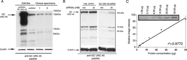
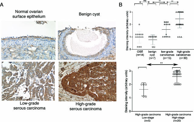
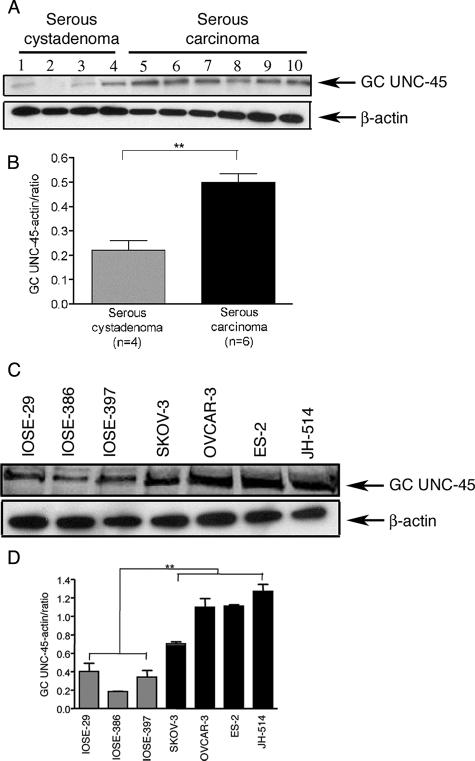
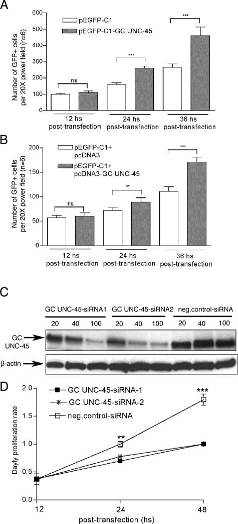
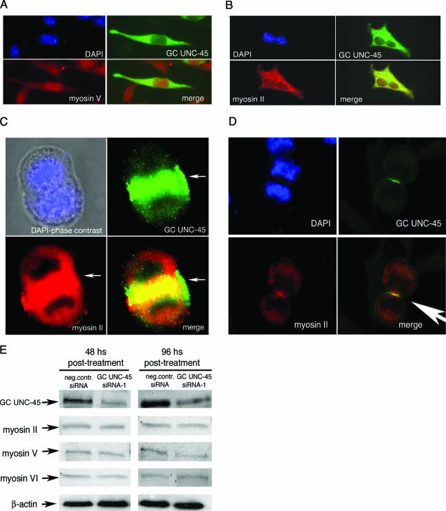
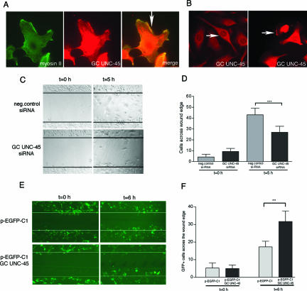
Both tumor cell proliferation and metastasis are dependent on myosin II. Because UNC-45 is required to chaperone the assembly of a functional myosin II motor, we examined the expression of the general cell (GC) UNC-45 isoform in ovarian tumors. Serous carcinoma expressed elevated levels of GC UNC-45 compared with normal ovarian surface epithelium and benign cystadenoma. High-stage exhibited greater GC UNC-45 expression than low-stage serous carcinoma. Similarly, GC UNC-45 transcripts and protein levels were higher in ovarian cell lines than in immortalized ovarian surface epithelial cells. Elevation of GC UNC-45 levels by ectopic expression enhanced the rate of ovarian cancer cell proliferation, whereas siRNA knockdown of GC UNC-45 suppressed proliferation without altering myosin II levels. GC UNC-45 and myosin II were diffuse within the cytoplasm of confluent interphase cells, but both accumulated together at the cleavage furrow during cytokinesis. GC UNC-45 and myosin II also trafficked to the leading edges of ovarian cancer cells induced to move in a scratch assay. Knockdown of GC UNC-45 reduced the spreading ability of ovarian cancer cells whereas it was enhanced by GC UNC-45 overexpression. In sum, these findings implicate elevated GC UNC-45 protein expression in ovarian carcinoma proliferation and metastasis.


Figures

Figure 1
Figure 1
GC UNC-45 antisera characterization. A: Affinity-purified antibodies to GC UNC-45 peptide were tested by Western blot analysis for reactivity to endogenous and ectopically expressed GC UNC-45 in the ES-2 ovarian cancer cell line as well as in clinical specimens of ovarian cancer. Equal loading was verified by using an antibody directed against β-actin. B: Rabbit antisera to GC UNC-45 peptide was tested by Western blot analysis for reactivity to endogenous GC UNC-45 in an ES-2 ovarian cancer cell line transfected with GC UNC-45 siRNA oligomer (GC UNC-45-siRNA-1) or with nonsilencing siRNA (neg. control-siRNA). GC UNC-45 expression level was tested by immunoblot analysis at 48 hours after transfection. Equal loading was verified by using an antibody directed against β-actin. C: Serial dilution of ES-2 cell lysate was immunoblotted for GC UNC-45. The density of immunoreactive bands was quantified by using Scion Image software using automatic background subtraction. The values were plotted and linear regression was calculated. Naphthalene Blue-Black-stained blot confirmed the loading of twofold dilutions of total cell lysate (not shown).
Figure 2
Figure 2
Immunohistochemical staining of GC UNC-45 in clinical specimens. A: Representative examples of intense GC UNC-45 staining in high-grade serous carcinoma, moderate staining intensity in low-grade serous carcinoma, and weak staining intensity in benign cyst and normal ovarian surface epithelium. B: Staining intensity for each case was graded as 0 (no staining), 1 (weak staining), 2 (moderate staining), and 3 (intense staining). **P < 0.02 statistical significance in staining intensities among indicated groups (Mann-Whitney U-test). Error bars indicate SD. Original magnifications: ×40.
Figure 3
Figure 3
Western blot analysis of GC UNC-45 in clinical specimens and cell lines. A: Lysates of serous cystadenoma (lanes 1 to 4) and serous carcinoma (lanes 5 to 10) were probed using GC UNC-45 peptide-specific rabbit antibody by Western blot. Equal loading was verified by using an antibody directed against β-actin. B: Ratio between GC UNC-45 and β-actin in tissue lysates from serous cystadenoma (four cases) and serous carcinoma (six cases). **P < 0.02. C: Lysates of immortalized ovarian surface epithelial cell lines (IOSE-29, IOSE-386, and IOSE-397) and ovarian cancer cell lines (SKOV-3, OVCAR-3, ES-2, and JH-514) were probed with anti-GC UNC-45 peptide rabbit antibody by Western blot. Equal loading was verified by using an antibody directed against β-actin. D: Ratio between GC UNC-45 and β-actin in tissue lysates from IOSEs and ovarian cancer cell lines, **P < 0.02. Error bars indicate SD.
Figure 4
Figure 4
GC UNC-45 level influences ovarian cancer cell proliferation. A: Proliferation rate of SKOV-3 cells ectopically expressing either GFP (pEGFP-C1 vector) alone or GFP-tagged GC UNC-45 (pEGFP-C1-GC UNC-45) was determined by counting the number of fluorescent cells 12, 24, and 36 hours after transfection. ***P < 0.001. B: Proliferation rate of SKOV-3 co-transfected with either pEGFP-C1 and pcDNA3 or pEGFP-C1 and pcDNA3-GC UNC-45 was determined by counting the number of fluorescent cells 12, 24, and 36 hours after transfection. ***P < 0.001. C: Efficiency of GC UNC-45 knockdown in ES-2 cells transfected with one of two different GC UNC-45 siRNA oligomers (GC UNC-45-siRNA-1 or -2) or with nonsilencing siRNA (neg. control-siRNA) at the indicated concentration was tested by immunoblot analysis at 48 hours after transfection. Equal loading was verified by using an antibody directed against β-actin. D: Proliferation rate of ES-2 cell line transfected with nonsilencing siRNA, GC UNC-45-siRNA-1, or GC UNC-45-siRNA-2 was measured by XTT assay starting from 12 hours after transfection. Results are expressed in terms of daily proliferation rate. **P < 0.02, ***P < 0.001. Error bars indicate SD.
Figure 5
Figure 5
Co-localization of GC UNC-45 with myosin II during cytokinesis and its effect on myosin expression levels. A: SKOV-3 cells were transfected with p-EGFP-C1-GC UNC-45 and stained with the DNA stain DAPI and anti-myosin V antibody. B: SKOV-3 cells were transfected with p-EGFP-C1-GC UNC-45 and stained with the DNA stain DAPI and anti-myosin II antibody. C: Mitotic SKOV-3 cells stained with the DNA stain DAPI, anti-GC UNC-45 peptide and anti-myosin II. A: The cellular localization of GC UNC-45 only partially overlaps myosin V. In particular, whereas significant myosin V staining is present over the nuclei, very little GC UNC-45 is present over the nuclei. In contrast, there is a significant and robust co-localization of GC UNC-45 with myosin II in interphase (B), metaphase (C), and telophase (D) of mitotic cells (indicated by the arrow pointing toward the cleavage furrow). E: Lysate of ES-2 cells transfected with negative control siRNA or GC UNC-45-siRNA-1 (100 nmol/L) for the time indicated was immunoblotted with antibodies against myosin II, myosin V, or myosin VI. Equal protein loading was verified by using an antibody directed against β-actin. Original magnifications, ×630 (D).
Figure 6
Figure 6
Localization of GC UNC-45 on the leading edges and its effect on cell motility. A: Microchambers of confluent SKOV-3 ovarian cancer cells were scratched, and migrating cells were stained with anti-myosin II and anti-GC UNC-45 peptide antibody 5 hours after scratch. Arrow indicates overlapping staining between myosin II and GC UNC-45 in the leading edges of moving cells. B: Confluent (left) and subconfluent (right) SKOV-3 ovarian cancer cell lines were stained with anti-GC UNC-45. Arrows indicate GC UNC-45 perinuclear localization in confluent cultures and GC UNC-45 localization in leading edges of spreading cells. C: Plates of confluent ES-2 cells were examined by phase-contrast microscopy at the time of removal by scratching (t = 0 hours) and 6 hours later (t = 6 hours). Fewer ES-2 cells transfected with GC UNC-45-siRNA spread across the plastic substratum compared with ES-2 cells transfected with neg.control-siRNA. D: Analysis of the number of cells spreading across the wound, results are means ± SD of three independent experiments. ***P < 0.001. E: Plates of confluent SKOV-3 p-EGFP-C1- or p-EGFP-C1-GC UNC-45-transfected cells were examined by immunofluorescent microscopy at the time of removal by scratching (t = 0 hours) and 6 hours later (t = 6 hours). SKOV-3 cells transfected with p-EGFP-C1-GC spread more rapidly across the plastic substratum into the wounded area compared with SKOV-3 p-EGFP-C1-transfected cells. F: Analysis of the number of cells spreading across the wound. Results are means ± SD of three independent experiments. **P < 0.02. Original magnifications, ×630 (B).

References

    1. Armstrong DK, Bundy B, Wenzel L, Huang HQ, Baergen R, Lele S, Copeland LJ, Walker JL, Burger RA. Intraperitoneal cisplatin and paclitaxel in ovarian cancer. N Engl J Med. 2006;354:34–43. - PubMed
    1. Bazzaro M, Lee MK, Zoso A, Stirling WL, Santillan A, Shih Ie M, Roden RB. Ubiquitin-proteasome system stress sensitizes ovarian cancer to proteasome inhibitor-induced apoptosis. Cancer Res. 2006;66:3754–3763. - PubMed
    1. Balasubramanian MK, Bi E, Glotzer M. Comparative analysis of cytokinesis in budding yeast, fission yeast and animal cells. Curr Biol. 2004;14:R806–R818. - PubMed
    1. Bresnick AR. Molecular mechanisms of nonmuscle myosin-II regulation. Curr Opin Cell Biol. 1999;11:26–33. - PubMed
    1. Edwards KA, Kiehart DP. Drosophila nonmuscle myosin II has multiple essential roles in imaginal disc and egg chamber morphogenesis. Development. 1996;122:1499–1511. - PubMed

Publication types