. 2007 Nov;189(22):8024-33.
doi: 10.1128/JB.01047-07. Epub 2007 Sep 14.

In vivo DNA binding of bacteriophage GA-1 protein p6

Affiliations

In vivo DNA binding of bacteriophage GA-1 protein p6

Martín Alcorlo et al. J Bacteriol. 2007 Nov.

Abstract

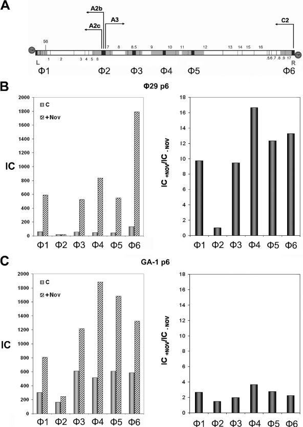
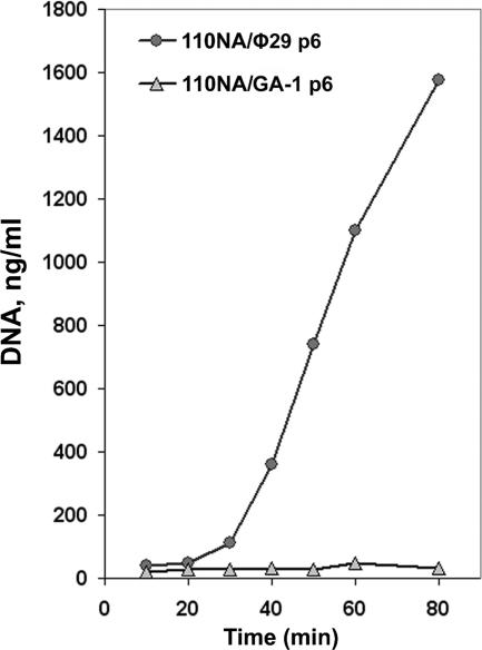
Bacteriophage GA-1 infects Bacillus sp. strain G1R and has a linear double-stranded DNA genome with a terminal protein covalently linked to its 5' ends. GA-1 protein p6 is very abundant in infected cells and binds DNA with no sequence specificity. We show here that it binds in vivo to the whole viral genome, as detected by cross-linking, chromatin immunoprecipitation, and real-time PCR analyses, and has the characteristics of a histone-like protein. Binding to DNA of GA-1 protein p6 shows little supercoiling dependency, in contrast to the ortholog protein of the evolutionary related Bacillus subtilis phage phi29. This feature is a property of the protein rather than the DNA or the cellular background, since phi29 protein p6 shows supercoiling-dependent binding to GA-1 DNA in Bacillus sp. strain G1R. GA-1 DNA replication is impaired in the presence of the gyrase inhibitors novobiocin and nalidixic acid, which indicates that, although noncovalently closed, the viral genome is topologically constrained in vivo. GA-1 protein p6 is also able to bind phi29 DNA in B. subtilis cells; however, as expected, the binding is less supercoiling dependent than the one observed with the phi29 protein p6. In addition, the nucleoprotein complex formed is not functional, since it is not able to transcomplement the DNA replication deficiency of a phi29 sus6 mutant. Furthermore, we took advantage of phi29 protein p6 binding to GA-1 DNA to find that the viral DNA ejection mechanism seems to take place, as in the case of phi29, with a right to left polarity in a two-step, push-pull process.


Figures

FIG. 1.
FIG. 1.
(A) Genetic and transcriptional map of the phage GA-1 linear DNA, showing the regions used to measure protein p6 binding by X-ChIP. Since DNA is sheared by sonication to an average size of ∼750 bp, each region would then comprise all of the overlapping DNA fragments that contain the amplified sequence (black boxes). According to the average size of the amplified sequences (∼300 bp) and that of the sonicated DNA (∼750 bp), every DNA region analyzed has ∼1,200 bp (gray boxes). This value would be only ∼750 bp for regions G1 and G5, since the amplified sequences are located at the ends of the linear DNA. The arrows point in the direction of transcription: the early promoters C2, A2b, and A2c transcribe leftward, and the late promoter A3 transcribes rightward. Genes that are conserved in comparison with phage φ29 are indicated with numbers. ORFs located at both genome ends that may encode several proteins, counterparts of which are not present in the genome of φ29, are indicated with letters. Circles represent the terminal protein attached to the 5′ DNA ends. L and R indicate the left and right ends of the GA-1 genome, respectively. (B) Protein p6 binding to GA-1 and plasmid DNA in the absence or presence of novobiocin and nalidixic acid. Bacillus sp. strain G1R cells harboring plasmid pPR55ow6 were grown, infected with GA-1 and, after 15 min, the untreated aliquot (C) was cross-linked and processed as described in Materials and Methods; the other two aliquots were treated with 34 μg of chloramphenicol/ml together with 500 μg of novobiocin/ml (+Nov) or nalidixic acid (+Nal) and cross-linked 10 min later. Protein p6 binding is expressed as the IC (see Materials and Methods). The IC values for each region are as follows (C, +Nov, +Nal): G1 = 4,864, 5,419, and 4,700; G2 = 3,242, 3,703, and 3,002; G3 = 2,659, 2,898, and 2,515; G4 = 3,123, 3,659, and 2,993; G5 = 4,553, 4,732, and 4,453; and P1 = 450, 511, and 420.
FIG. 2.
FIG. 2.
Effect of novobiocin and nalidixic acid on GA-1 DNA replication. Bacillus sp. strain G1R cells were infected with phage GA-1, and 25 min later the culture was divided into three aliquots and further grown in the presence of novobiocin (+Nov) or nalidixic acid (+Nal) (500 μg of each/ml) or with no addition (C). Aliquots were obtained at the indicated times after infection, and the DNA was purified by phenol extraction and ethanol precipitation. The amount of GA-1 DNA was calculated by real-time PCR of the left terminal sequence (L; see Fig. 1A). The data are expressed as nanograms of full-length GA-1 DNA per milliliter of culture.
FIG. 3.
FIG. 3.
φ29 protein p6 binding to GA-1 right terminus (G5) and plasmid region P1 in Bacillus sp. strain G1R. Cells harboring plasmid pPR55w6 were grown, and the culture was treated with 34 μg of chloramphenicol/ml and infected with phage GA-1. The culture was divided into two aliquots and, after 20 min, one aliquot was cross-linked (C), and the other was treated with 500 μg of novobiocin/ml (+Nov) and cross-linked 10 min later. φ29 protein p6 binding is expressed as the IC. (A) IC values for regions G1 and G5 (GA-1 left and right genome ends, respectively) are as follows: G1, not detected (ND); G5, 25.8 (C) and 130 (+Nov). (B) IC values for plasmid region P1 are as follows: 89.4 (C) and 911.9 (+Nov). The binding increase in the presence of novobiocin, expressed as the IC+Nov/IC−Nov ratio, is shown in the right panel.
FIG. 4.
FIG. 4.
(A) φ29 protein p6 binding to the φ29 right terminus (φ6) and plasmid region P1. B. subtilis 110NA cells producing φ29 protein p6 were grown and treated with 34 μg of chloramphenicol/ml together with or without 500 μg of novobiocin/ml. Cells were infected with φ29 sus14(1242) and cross-linked 40 min later. IC values for each DNA region in the absence or presence of novobiocin are shown in the left panel. The binding increase in the presence of novobiocin, expressed as the IC+Nov/IC−Nov ratio, is shown in the right panel. (B) GA-1 protein p6 binding to the φ29 right terminus (φ6) and plasmid region P1. GA-1 protein p6 synthesis was induced in B. subtilis 110NA cells as described in Materials and Methods. Cells processed as before were infected with φ29 sus14(1242) and cross-linked 40 min later. IC values in the absence or presence of novobiocin are shown in the left panel. The binding increase in the presence of novobiocin, expressed as the IC+Nov/IC−Nov ratio, is shown in the right panel.
FIG. 5.
FIG. 5.
(A) Genetic and transcriptional map of phage φ29 linear DNA. The sequences selected for PCR amplification are shown in black and the regions analyzed for protein p6 binding, φ1-φ6, are shown in gray, as explained in Fig. 1A. The position of genes 1 to 17 and 56 is indicated. .5 to .9 stands for 16.5 to 16.9, respectively. Circles represent the terminal protein attached to the 5′ DNA ends. L and R indicate the left and right ends of the φ29 genome, respectively. The arrows point the direction of transcription: the early promoters C2, A2b, and A2c leftward and the late promoter A3 rightward. (B) φ29 protein p6 binding to φ29 DNA. B. subtilis 110NA cells producing φ29 protein p6 were infected with φ29 sus6(626) and, 20 min later, an aliquot (c) was cross-linked as described in Materials and Methods; another aliquot (+Nov) was treated with 34 μg of chloramphenicol/ml together with 500 μg of novobiocin/ml and cross-linked 40 min later. Protein p6 binding for each region is shown in the left panel. The IC values (c and +Nov) are as follows: φ1, 60.5 and 590; φ2, 18.5 and 18.6; φ3, 55.8 and 527.7; φ4, 50.1 and 834.4; φ5, 44.4 and 548.5; φ6, 134.9 and 1791; and P1, 205.9 and 767.5. Binding increase for each φ29 DNA region in the presence of novobiocin, expressed as IC+Nov/IC−Nov ratio, is shown in the right panel. (C) GA-1 protein p6 binding to φ29 DNA. B. subtilis 110NA cells producing GA-1 protein p6 were infected with φ29 sus6(626) and aliquots (c) and (+Nov) were obtained as described above. Protein p6 binding for each region is shown in the left panel. The IC values (c and +Nov) are the following: φ1, 303.7 and 805.9; φ2, 162.9 and 245.3; φ3, 613.4 and 1,216; φ4, 515.7 and 1,885.5; φ5, 607.3 and 1,681.8; φ6, 585.4 and 1,325; and P1, 586.3 and 975. The binding increase for each φ29 DNA region in the presence of novobiocin, expressed as the IC+Nov/IC−Nov ratio, is shown in the right panel.
FIG. 6.
FIG. 6.
Transcomplementation of φ29 DNA synthesis in B. subtilis 110NA cells producing φ29 or GA-1 protein p6. B. subtilis 110NA cells producing φ29 or GA-1 protein p6 were infected with φ29 sus6(626), aliquots were taken at the indicated times after infection, and the DNA was purified by phenol extraction and ethanol precipitation. The amount of φ29 DNA was measured by real-time PCR of the left terminal sequence (L). The data are expressed as nanograms of full-length φ29 DNA per milliliter of culture.

Similar articles

Cited by

References

    1. Abril, A. M., S. Marco, J. L. Carrascosa, M. Salas, and J. M. Hermoso. 1999. Oligomeric structures of the phage φ29 histone-like protein p6. J. Mol. Biol. 292:581-588. - PubMed
    1. Abril, A. M., M. Salas, J. M. Andreu, J. M. Hermoso, and G. Rivas. 1997. Phage φ29 protein p6 is in a monomer-dimer equilibrium that shifts to higher association states at the millimolar concentrations found in vivo. Biochemistry 36:11901-11908. - PubMed
    1. Alcorlo, M., V. González-Huici, J. M. Hermoso, W. J. Meijer, and M. Salas. 2007. The phage φ29 membrane protein p16.7, involved in DNA replication, is required for efficient ejection of the viral genome. J. Bacteriol. 189:5542-5549. - PMC - PubMed
    1. Arwert, F., and G. Venema. 1974. Protease-sensitive transfection of Bacillus subtilis with bacteriophage GA-1 DNA: a probable case of heterologous transfection. J. Virol. 13:584-589. - PMC - PubMed
    1. Barthelemy, I., and M. Salas. 1989. Characterization of a new prokaryotic transcriptional activator and its DNA recognition site. J. Mol. Biol. 208:225-232. - PubMed

Publication types