. 2007 Sep 25;104(39):15311-6.
doi: 10.1073/pnas.0707190104. Epub 2007 Sep 14.

Crystallographic trapping in the rebeccamycin biosynthetic enzyme RebC

Affiliations

Crystallographic trapping in the rebeccamycin biosynthetic enzyme RebC

Katherine S Ryan et al. Proc Natl Acad Sci U S A. .

Abstract

The biosynthesis of rebeccamycin, an antitumor compound, involves the remarkable eight-electron oxidation of chlorinated chromopyrrolic acid. Although one rebeccamycin biosynthetic enzyme is capable of generating low levels of the eight-electron oxidation product on its own, a second protein, RebC, is required to accelerate product formation and eliminate side reactions. However, the mode of action of RebC was largely unknown. Using crystallography, we have determined a likely function for RebC as a flavin hydroxylase, captured two snapshots of its dynamic catalytic cycle, and trapped a reactive molecule, a putative substrate, in its binding pocket. These studies strongly suggest that the role of RebC is to sequester a reactive intermediate produced by its partner protein and to react with it enzymatically, preventing its conversion to a suite of degradation products that includes, at low levels, the desired product.


Conflict of interest statement

The authors declare no conflict of interest.

Figures

Fig. 1.
Fig. 1.
Structures of small molecules and overall reaction scheme. (a) Structures of compounds discussed in the article are shown schematically. (b) StaP (or RebP), incubated with an electron source, reacts with chromopyrrolic acid (4) to give three products, as shown. Addition of RebC leads to near-exclusive formation of arcyriaflavin A (5).
Fig. 2.
Fig. 2.
RebC structural comparisons. (a) The structure of native RebC is shown with domain I (red), domain II (blue), domain III (green), and a flavin in the OUT conformation (green carbons). Density for residues 354–363 are missing in this structure; two arrows indicate the residues bordering the missing density. (b) The structure of chromopyrrolic acid-soaked RebC, also colored by domain, shows flavin in the IN conformation (green carbons), with Fo-Fc omit density for the molecule captured in the substrate-binding pocket contoured at +3 σ. The “melting” helix, which traverses from domain I to II, is visible in this structure, and two arrows indicate the residues bordering this helix. (c) Chromopyrrolic acid-soaked RebC (gray) is shown with aligned portions of para-hydroxybenzoate hydroxylase (pale blue, 1DOC, 248 of 394 residues), phenol hydroxylase (pale pink, 1PN0C, 281 of 664 residues), and meta-hydroxybenzoate hydroxylase (pale yellow, 2DKH, 264 of 639 residues). Aligned FAD molecules, which have been observed in the IN conformation, are shown in black (RebC), blue (para-hydroxybenzoate hydroxylase), and red (phenol hydroxylase).
Fig. 3.
Fig. 3.
Reduction potential of RebC. The redox potential of RebC was determined by using the method of Massey (18) using 1-hydroxyphenazine (E°′ = −172 mV) as a reference dye (see SI Methods).
Fig. 4.
Fig. 4.
Mobile flavins. (a) Phenol hydroxylase (PDB ID code 1PN0) (12) has a mobile flavin, as seen from the superimposed structures of the FAD, which is found in both the OUT (light blue carbons) and IN (green carbons) conformations. The substrate phenol is also shown. (b) Two positions of FAD in RebC, manually superimposed from structures of native (light blue carbons, OUT) and chromopyrrolic acid-soaked structures (green carbons, IN), are shown. The 7-carboxy-K252c, which was refined into the chromopyrrolic acid-soaked structure, is shown with green carbons. (c) Cut-away view of FAD (light blue carbons) with a 2Fo-Fc omit map at 1σ shows that the flavin in the native structure is OUT, with a hydrogen bond between the isoalloxazine ring and Arg-239. The isoalloxazine ring is stacked between Arg-46 and Trp-276. Because FAD is thought to be reduced by NAD(P)H in the OUT conformation via a reaction at the re face of the isoalloxazine ring (23), Trp-276 would have to move aside for reduction to occur, just as proposed for the structurally homologous residue, Tyr-317 in meta-hydroxybenzoate hydroxylase, which also stacks on the re face of its isoalloxazine ring (13). (d) Cut-away view of FAD (green carbons) in the chromopyrrolic acid-soaked structure shows that the isoalloxazine ring in the IN conformation no longer stacks between Trp-276 and Arg-46, and the interaction with Arg-239 is lost. Arg-46 now interacts with FAD phosphate oxygens, and Arg-239 interacts with bound 7-carboxy-K252c (green carbons). The 2Fo-Fc omit map is contoured at 1 σ.
Fig. 5.
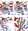
Fig. 5.
Substrate-binding site. (a) The 7-carboxy-K252c, which was refined in the chromopyrrolic acid-soaked structure, is bound at the interface of domain I (red) and II (blue), surrounded by a number of hydrophobic side chains, including Phe-227, which shifts conformation. Leu-358 is found on the helix that is disordered in the native structure. A loop (red, residues 303–306) from domain I interacts with one face of 7-carboxy-K252c. Hydrogen-bonding interactions with 7-carboxy-K252c include Glu-396, Arg-230, and Arg-239. An Fo-Fc omit map is contoured at 3.0 σ. (b) Binding site of the K252c-soaked structure shows the isoalloxazine ring of flavin (green) locked in an OUT conformation. Trp-276 stacks on the re face of the isoalloxazine ring, Phe-227 is not shifted, and Arg-230 is too far to interact with K252c. Charged residue Glu-396 hydrogen-bonds with the indole nitrogens, and Arg-239 hydrogen-bonds with the FAD, and it does not interact with the substrate. K252c is contoured with an Fo-Fc omit map at 3.0 σ. (c) The native structure of RebC shows the exposure of the substrate-binding pocket to solvent with the absence of the melting helix. Additionally, Glu-396 is disordered, Phe-227 is not shifted, and Phe-216 assumes two alternate conformations. FAD (green), as in the K252c-soaked structure, is positioned in the OUT conformation. (d) Cut-away view of 7-carboxy-K252c and FAD-binding site shows that one ring carbon of 7-carboxy-K252c is 5.1 Å from the C4a carbon of FAD. One oxygen of the carboxylate of 7-carboxy-K252c additionally has hydrogen bonds to both Arg-230 and Arg-239.

Comment in

  • Crystallography gets the jump on the enzymologists.
    Ballou DP. Ballou DP. Proc Natl Acad Sci U S A. 2007 Oct 2;104(40):15587-8. doi: 10.1073/pnas.0707843104. Epub 2007 Sep 26. Proc Natl Acad Sci U S A. 2007. PMID: 17898164 Free PMC article. No abstract available.

References

    1. Bush JA, Long BH, Catino JJ, Bradner WT, Tomita K. J Antibiot. 1987;40:668–678. - PubMed
    1. Staker BL, Feese MD, Cushman M, Pommier Y, Zembower D, Stewart L, Burgin AB. J Med Chem. 2005;48:2336–2345. - PubMed
    1. Prudhomme M. Eur J Med Chem. 2003;38:123–140. - PubMed
    1. Sanchez C, Butovich IA, Brana AF, Rohr J, Mendez C, Salas JA. Chem Biol. 2002;9:519–531. - PubMed
    1. Onaka H, Taniguchi S, Igarashi Y, Furumai T. Biosci Biotech Biochem. 2003;67:127–138. - PubMed

Publication types

LinkOut - more resources