. 2007 Sep 25;104(39):15508-13.
doi: 10.1073/pnas.0706532104. Epub 2007 Sep 14.

Type VI secretion system translocates a phage tail spike-like protein into target cells where it cross-links actin

Affiliations

Type VI secretion system translocates a phage tail spike-like protein into target cells where it cross-links actin

Stefan Pukatzki et al. Proc Natl Acad Sci U S A. .

Abstract

Genes encoding type VI secretion systems (T6SS) are widely distributed in pathogenic Gram-negative bacterial species. In Vibrio cholerae, T6SS have been found to secrete three related proteins extracellularly, VgrG-1, VgrG-2, and VgrG-3. VgrG-1 can covalently cross-link actin in vitro, and this activity was used to demonstrate that V. cholerae can translocate VgrG-1 into macrophages by a T6SS-dependent mechanism. Protein structure search algorithms predict that VgrG-related proteins likely assemble into a trimeric complex that is analogous to that formed by the two trimeric proteins gp27 and gp5 that make up the baseplate "tail spike" of Escherichia coli bacteriophage T4. VgrG-1 was shown to interact with itself, VgrG-2, and VgrG-3, suggesting that such a complex does form. Because the phage tail spike protein complex acts as a membrane-penetrating structure as well as a conduit for the passage of DNA into phage-infected cells, we propose that the VgrG components of the T6SS apparatus may assemble a "cell-puncturing device" analogous to phage tail spikes to deliver effector protein domains through membranes of target host cells.


Conflict of interest statement

The authors declare no conflict of interest.

Figures

Fig. 1.
Fig. 1.
Hcp export requires the presence of VgrG-1 and VgrG-2. Plasmids allowing the inducible expression of HA-tagged VgrG-1 (pVgrG1), vsvG-tagged VgrG-2 (pVgrG2), or plasmid control (control) were introduced into wild-type V. cholerae, or isogenic mutants lacking VgrG-1 (ΔvgrG1) or VgrG-2 (ΔvgrG2). Culture supernatants from cells grown in liquid broth under inducing (+) and noninducing (−) conditions were concentrated and separated by SDS/PAGE; Hcp was visualized by Western blot analysis with Hcp antiserum. To guarantee plasmid-borne expression of HA-tagged VgrG-1 and vsvG-tagged VgrG-2, cell extracts were separated by SDS/PAGE for immunoblotting with HA and vsvG antisera, respectively.
Fig. 2.
Fig. 2.
Recombinant VgrG-1 cross-links actin in an ATP-dependent fashion. (A) Hexahistidine-tagged VgrG-1 was purified on a nickel-NTA column and separated by SDS/PAGE (see arrow). (B) Recombinant VgrG-1 was incubated at 37°C for 2 h with purified rabbit skeletal monomeric G-actin (Cytoskeleton, Denver, CO) or crude lysates from Dictyostelium and RAW 264.7. Reactions were supplemented with 2 mM Mg-ATP as indicated. Reaction mixtures were separated by SDS/PAGE, and actin was visualized by Western blot analysis with actin antiserum.
Fig. 3.
Fig. 3.
VgrG-1-mediated cross-linking of the host actin cytoskeleton. Cultured J774 cells were infected for 2 h with indicated V. cholerae strains at a multiplicity of infection (MOI) of 10. Extracts of infected cells were separated by SDS/PAGE for immunoblotting with actin antiserum.
Fig. 4.
Fig. 4.
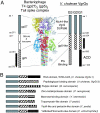
VgrGs share structural features with the phage tail of bacteriophage T4. (A) V. cholera VgrGs are a fusion of the phage tail proteins gp27 (gray) and gp5 (hatched), omitting the OB-fold and lysozyme domains (white) of the phage gp5 protein. The gp5 domain consists of a β-helix repeat (black arrows) that forms the cell-puncturing “needle” of the phage tail. VgrG-1 and VgrG-3 contain a C-terminal actin-cross-linking domain (ACD) and peptidoglycan (PG)-binding domain, respectively. Amino acid numbers are designated for gp27, gp5, and VgrG-1. Crystal structure for the phage tail spike (gp27 and gp5) are shown for reference (24) (adapted by permission from Macmillan Publishers Ltd: Nature, 31 January 2002). (B) Graphical representation of a subset of the VgrG-family members with extended C-terminal domains. These proteins consist of a gp27-like domain (solid black), gp5-like domain (hatched) and different C-terminal extensions (bars with various patterns). On the right is shown the predicted structural similarities of the C-terminal domains identified by the PSIPRED protein structure prediction server (http://bioinf.cs.ucl.ac.uk/psipred/). Protein structures schematically shown from top to bottom have the following accession numbers: AAF94573, AAF96037, ZP_00981404, NP_248953, ZP_00832947, CAC92964, YP_652030, and YP_606302. A more extensive list of VgrG orthologs with extended C termini together with PSIPRED predicted structural similarities can be found in SI Table 1.
Fig. 5.
Fig. 5.
Immunoprecipitation of VgrG-1-myc. V. cholerae strains expressing either wild-type VgrG-1 or a VgrG-1::myc tagged protein were grown to midlogarithmic growth phase. Culture supernatants were filtered and Myc antisera conjugated to agarose beads were added to pull-down VgrG-1::myc and any interacting proteins. Resulting precipitates were separated by SDS/PAGE for immunoblotting with antisera specific for VgrG-1, VgrG-2, and VgrG-3. The bands corresponding to the size of VgrG-1, VgrG-2, and VgrG-3 were identified only in immunoprecipitations from supernatants derived from the strain expressing VgrG-1-myc.

Similar articles

Cited by

References

    1. Gerlach RG, Hensel M. Int J Med Microbiol. 2007 doi: 10.1016/j.ijmm.2007.03.017. - DOI - PubMed
    1. Johnson TL, Abendroth J, Hol WG, Sandkvist M. FEMS Microbiol Lett. 2006;255:175–186. - PubMed
    1. Rosqvist R, Hakansson S, Forsberg A, Wolf-Watz H. EMBO J. 1995;14:4187–4195. - PMC - PubMed
    1. Kirby JE, Isberg RR. Trends Microbiol. 1998;6:256–258. - PubMed
    1. Segal G, Shuman HA. Trends Microbiol. 1998;6:253–255. - PubMed

Publication types