

Unraveling the difference between invertases and fructan exohydrolases: a single amino acid (Asp-239) substitution transforms Arabidopsis cell wall invertase1 into a fructan 1-exohydrolase
- PMID: 17873089
- PMCID: PMC2048769
- DOI: 10.1104/pp.107.105049
Unraveling the difference between invertases and fructan exohydrolases: a single amino acid (Asp-239) substitution transforms Arabidopsis cell wall invertase1 into a fructan 1-exohydrolase
Abstract
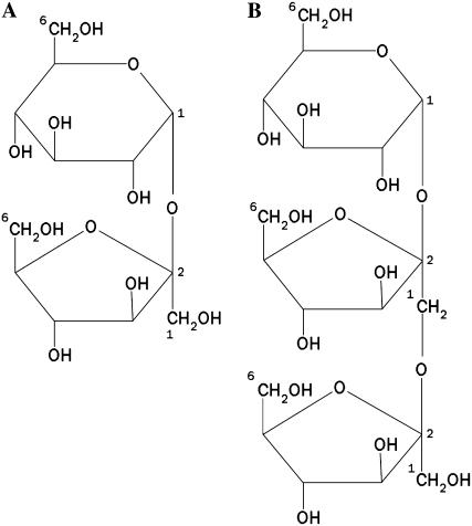
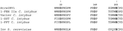
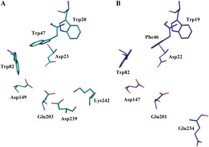
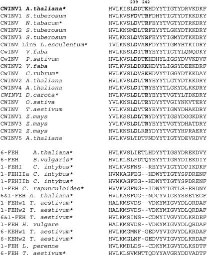
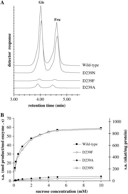
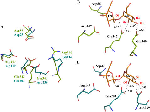
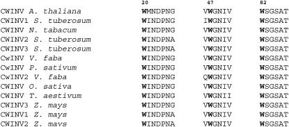
Plant cell wall invertases and fructan exohydrolases (FEHs) are very closely related enzymes at the molecular and structural level (family 32 of glycoside hydrolases), but they are functionally different and are believed to fulfill distinct roles in plants. Invertases preferentially hydrolyze the glucose (Glc)-fructose (Fru) linkage in sucrose (Suc), whereas plant FEHs have no invertase activity and only split terminal Fru-Fru linkages in fructans. Recently, the three-dimensional structures of Arabidopsis (Arabidopsis thaliana) cell wall Invertase1 (AtcwINV1) and chicory (Cichorium intybus) 1-FEH IIa were resolved. Until now, it remained unknown which amino acid residues determine whether Suc or fructan is used as a donor substrate in the hydrolysis reaction of the glycosidic bond. In this article, we present site-directed mutagenesis-based data on AtcwINV1 showing that the aspartate (Asp)-239 residue fulfills an important role in both binding and hydrolysis of Suc. Moreover, it was found that the presence of a hydrophobic zone at the rim of the active site is important for optimal and stable binding of Suc. Surprisingly, a D239A mutant acted as a 1-FEH, preferentially degrading 1-kestose, indicating that plant FEHs lacking invertase activity could have evolved from a cell wall invertase-type ancestor by a few mutational changes. In general, family 32 and 68 enzymes containing an Asp-239 functional homolog have Suc as a preferential substrate, whereas enzymes lacking this homolog use fructans as a donor substrate. The presence or absence of such an Asp-239 homolog is proposed as a reliable determinant to discriminate between real invertases and defective invertases/FEHs.
Figures
Similar articles
-
N-glycosylation affects substrate specificity of chicory fructan 1-exohydrolase: evidence for the presence of an inulin binding cleft.New Phytol. 2007;176(2):317-324. doi: 10.1111/j.1469-8137.2007.02174.x. New Phytol. 2007. PMID: 17888113
-
Donor and acceptor substrate selectivity among plant glycoside hydrolase family 32 enzymes.FEBS J. 2009 Oct;276(20):5788-98. doi: 10.1111/j.1742-4658.2009.07316.x. Epub 2009 Sep 17. FEBS J. 2009. PMID: 19765078 Review.
-
Influencing the binding configuration of sucrose in the active sites of chicory fructan 1-exohydrolase and sugar beet fructan 6-exohydrolase.New Phytol. 2008;178(3):572-80. doi: 10.1111/j.1469-8137.2008.02386.x. Epub 2008 Mar 3. New Phytol. 2008. PMID: 18331426
-
Unexpected presence of fructan 6-exohydrolases (6-FEHs) in non-fructan plants: characterization, cloning, mass mapping and functional analysis of a novel "cell-wall invertase-like" specific 6-FEH from sugar beet (Beta vulgaris L.).Plant J. 2003 Dec;36(5):697-710. doi: 10.1046/j.1365-313x.2003.01912.x. Plant J. 2003. PMID: 14617070
-
Structural insights into glycoside hydrolase family 32 and 68 enzymes: functional implications.J Exp Bot. 2009;60(3):727-40. doi: 10.1093/jxb/ern333. Epub 2009 Jan 6. J Exp Bot. 2009. PMID: 19129163 Review.
Cited by
-
The Important Roles Played in Substrate Binding of Aromatic Amino Acids in Exo-Inulinase From Kluyveromyces cicerisporus CBS 4857.Front Mol Biosci. 2020 Sep 25;7:569797. doi: 10.3389/fmolb.2020.569797. eCollection 2020. Front Mol Biosci. 2020. PMID: 33102520 Free PMC article.
-
Genome-Wide Survey of Invertase Encoding Genes and Functional Characterization of an Extracellular Fungal Pathogen-Responsive Invertase in Glycine max.Int J Mol Sci. 2018 Aug 14;19(8):2395. doi: 10.3390/ijms19082395. Int J Mol Sci. 2018. PMID: 30110937 Free PMC article.
-
Cloning and functional characterization of a fructan 1-exohydrolase (1-FEH) in the cold tolerant Patagonian species Bromus pictus.Planta. 2009 Dec;231(1):13-25. doi: 10.1007/s00425-009-1020-5. Epub 2009 Sep 30. Planta. 2009. PMID: 19789892
-
Pp6-FEH1 encodes an enzyme for degradation of highly polymerized levan and is transcriptionally induced by defoliation in timothy (Phleum pratense L.).J Exp Bot. 2011 Jun;62(10):3421-31. doi: 10.1093/jxb/err018. Epub 2011 Feb 11. J Exp Bot. 2011. PMID: 21317211 Free PMC article.
-
Metabolome and transcriptome analyses reveal changes of rapeseed in response to ABA signal during early seedling development.BMC Plant Biol. 2024 Apr 5;24(1):245. doi: 10.1186/s12870-024-04918-8. BMC Plant Biol. 2024. PMID: 38575879 Free PMC article.
References
-
- Alberto F, Bignon C, Sulzenbacher G, Henrissat B, Czjzek M (2004) The three-dimensional structure of invertase (beta-fructosidase) from Thermotoga maritima reveals a bimodular arrangement and an evolutionary relationship between retaining and inverting glycosidases. J Biol Chem 279 18903–18910 - PubMed
-
- Altenbach D, Nüesch E, Ritsema T, Boller T, Wiemken A (2005) Mutational analysis of the active center of plant fructosyltransferases: Festuca 1-SST and barley 6-SFT. FEBS Lett 579 4647–4653 - PubMed
Publication types
MeSH terms
Substances
LinkOut - more resources
Full Text Sources
Other Literature Sources
Molecular Biology Databases
