

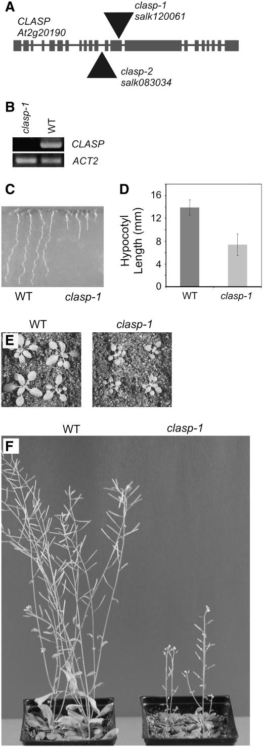
The Arabidopsis CLASP gene encodes a microtubule-associated protein involved in cell expansion and division
- PMID: 17873093
- PMCID: PMC2048705
- DOI: 10.1105/tpc.107.053777
The Arabidopsis CLASP gene encodes a microtubule-associated protein involved in cell expansion and division
Abstract
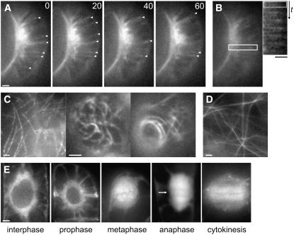
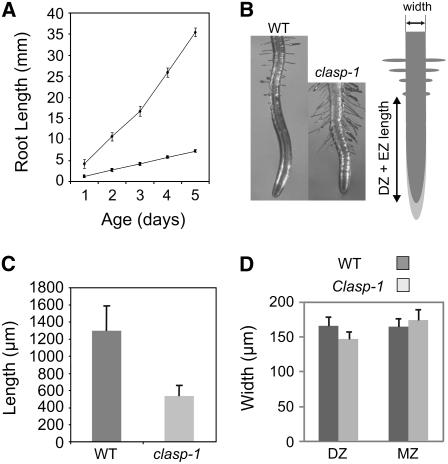
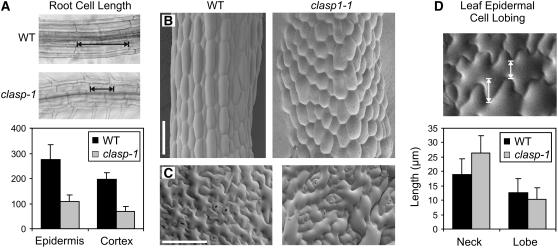
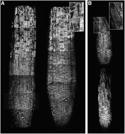
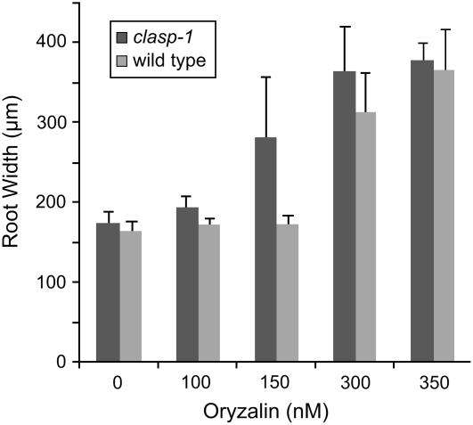
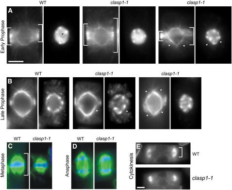
Controlling microtubule dynamics and spatial organization is a fundamental requirement of eukaryotic cell function. Members of the ORBIT/MAST/CLASP family of microtubule-associated proteins associate with the plus ends of microtubules, where they promote the addition of tubulin subunits into attached kinetochore fibers during mitosis and stabilize microtubules in the vicinity of the plasma membrane during interphase. To date, nothing is known about their function in plants. Here, we show that the Arabidopsis thaliana CLASP protein is a microtubule-associated protein that is involved in both cell division and cell expansion. Green fluorescent protein-CLASP localizes along the full length of microtubules and shows enrichment at growing plus ends. Our analysis suggests that CLASP promotes microtubule stability. clasp-1 T-DNA insertion mutants are hypersensitive to microtubule-destabilizing drugs and exhibit more sparsely populated, yet well ordered, root cortical microtubule arrays. Overexpression of CLASP promotes microtubule bundles that are resistant to depolymerization with oryzalin. Furthermore, clasp-1 mutants have aberrant microtubule preprophase bands, mitotic spindles, and phragmoplasts, indicating a role for At CLASP in stabilizing mitotic arrays. clasp-1 plants are dwarf, have significantly reduced cell numbers in the root division zone, and have defects in directional cell expansion. We discuss possible mechanisms of CLASP function in higher plants.
Figures

References
-
- Abe, T., and Hashimoto, T. (2005). Altered microtubule dynamics by expression of modified alpha-tubulin protein causes right-handed helical growth in transgenic Arabidopsis plants. Plant J. 43 191–204. - PubMed
-
- Abe, T., Thitamadee, S., and Hashimoto, T. (2004). Microtubule defects and cell morphogenesis in the lefty1lefty2 tubulin mutant of Arabidopsis thaliana. Plant Cell Physiol. 45 211–220. - PubMed
-
- Akhmanova, A., and Hoogenraad, C.C. (2005). Microtubule plus-end-tracking proteins: Mechanisms and functions. Curr. Opin. Cell Biol. 17 47–54. - PubMed
-
- Akhmanova, A., Hoogenraad, C.C., Drabek, K., Stepanova, T., Dortland, B., Verkerk, T., Vermeulen, W., Burgering, B.M., De Zeeuw, C.I., Grosveld, F., and Galjart, N. (2001). Clasps are CLIP-115 and -170 associating proteins involved in the regional regulation of microtubule dynamics in motile fibroblasts. Cell 104 923–935. - PubMed
Publication types
MeSH terms
Substances
LinkOut - more resources
Full Text Sources
Other Literature Sources
Molecular Biology Databases
