Comparative Study
. 2007 Nov;85(2):133-48.
doi: 10.1007/s11060-007-9400-9. Epub 2007 Sep 15.

Intracranial glioblastoma models in preclinical neuro-oncology: neuropathological characterization and tumor progression

Affiliations
Comparative Study

Intracranial glioblastoma models in preclinical neuro-oncology: neuropathological characterization and tumor progression

Marianela Candolfi et al. J Neurooncol. 2007 Nov.

Abstract

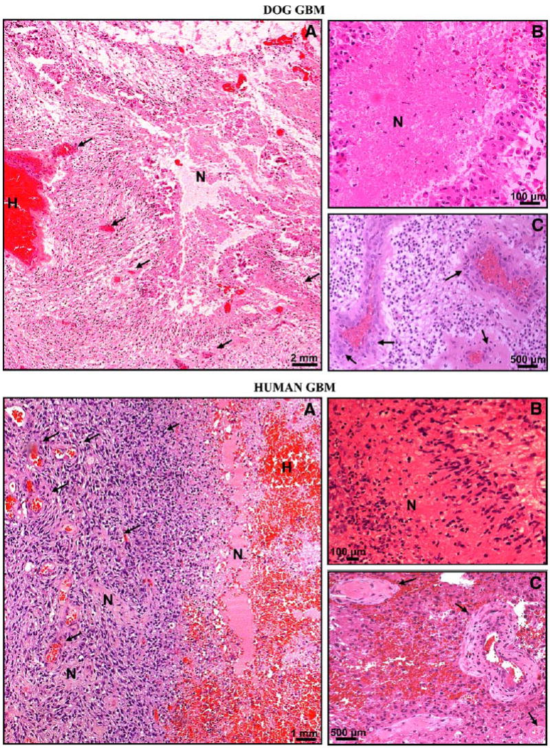
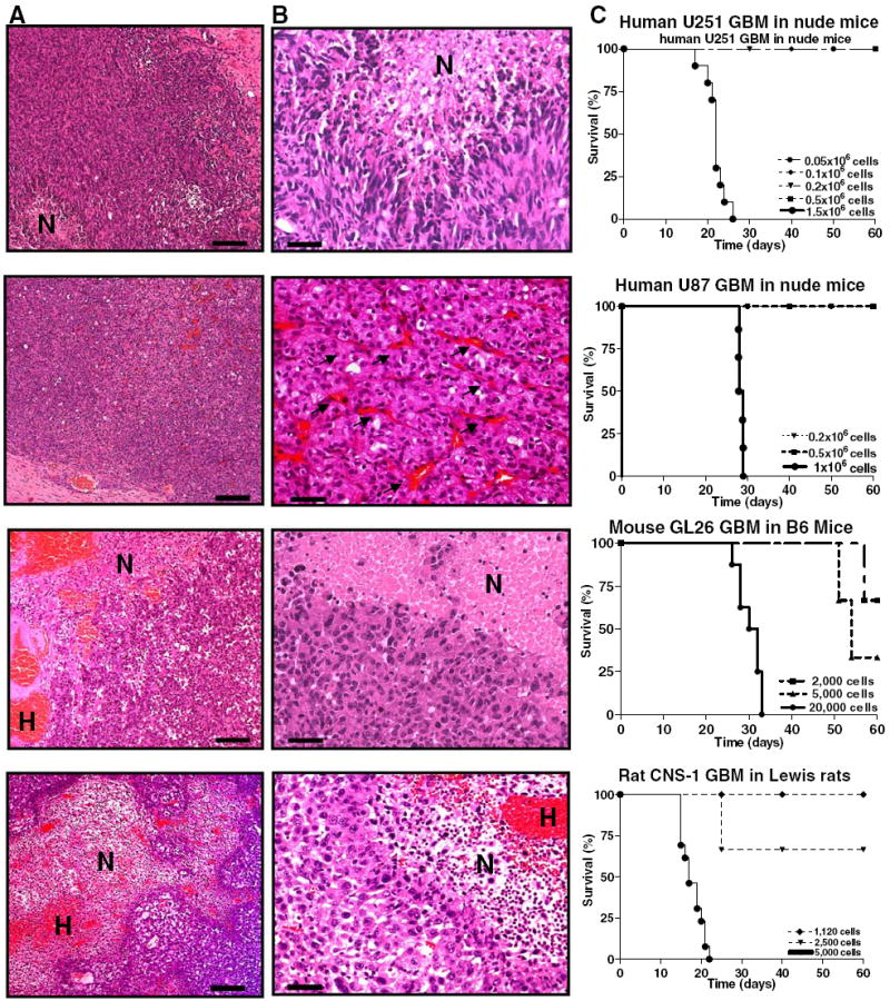
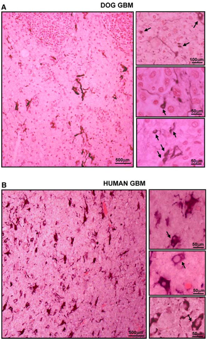
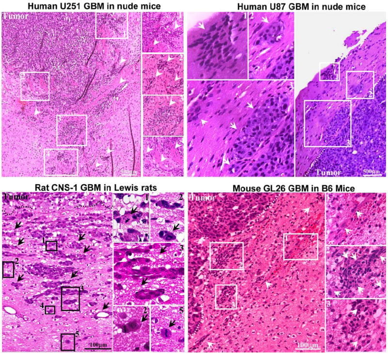
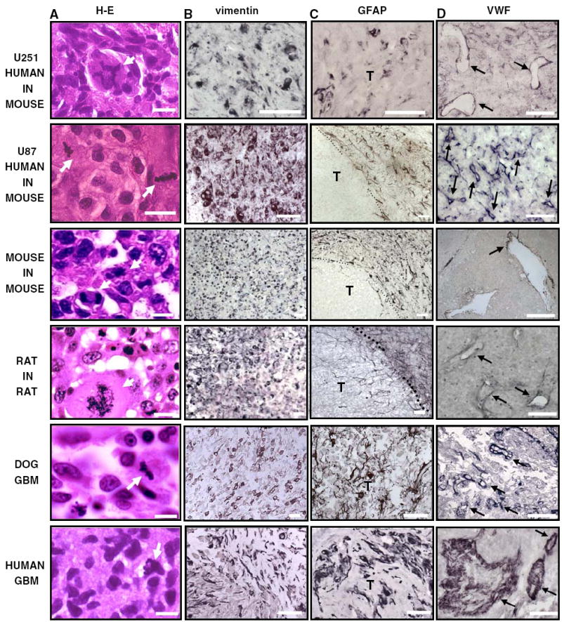
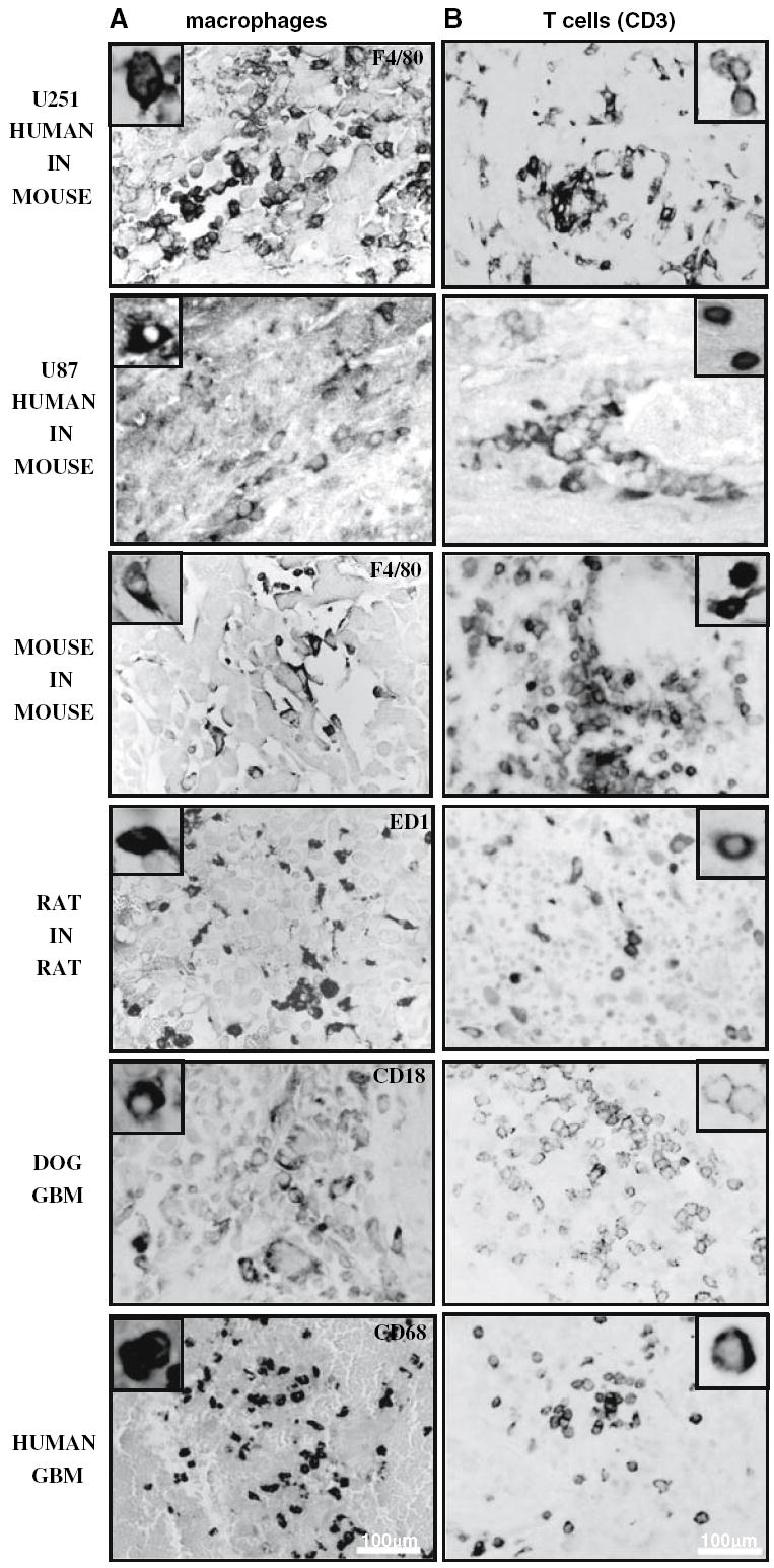
Although rodent glioblastoma (GBM) models have been used for over 30 years, the extent to which they recapitulate the characteristics encountered in human GBMs remains controversial. We studied the histopathological features of dog GBM and human xenograft GBM models in immune-deficient mice (U251 and U87 GBM in nude Balb/c), and syngeneic GBMs in immune-competent rodents (GL26 cells in C57BL/6 mice, CNS-1 cells in Lewis rats). All GBMs studied exhibited neovascularization, pleomorphism, vimentin immunoreactivity, and infiltration of T-cells and macrophages. All the tumors showed necrosis and hemorrhages, except the U87 human xenograft, in which the most salient feature was its profuse neovascularization. The tumors differed in the expression of astrocytic intermediate filaments: human and dog GBMs, as well as U251 xenografts expressed glial fibrillary acidic protein (GFAP) and vimentin, while the U87 xenograft and the syngeneic rodent GBMs were GFAP(-) and vimentin(+). Also, only dog GBMs exhibited endothelial proliferation, a key feature that was absent in the murine models. In all spontaneous and implanted GBMs we found histopathological features compatible with tumor invasion into the non-neoplastic brain parenchyma. Our data indicate that murine models of GBM appear to recapitulate several of the human GBM histopathological features and, considering their reproducibility and availability, they constitute a valuable in vivo system for preclinical studies. Importantly, our results indicate that dog GBM emerges as an attractive animal model for testing novel therapies in a spontaneous tumor in the context of a larger brain.


Figures

Fig. 1
Fig. 1
Histopathological comparisons between dog (top panels) and human (bottom panels) spontaneous GBM. Paraffin sections (5 μm) of spontaneous human and canine GBM were stained with hematoxylin and eosin (H–E) to determine their histopathologic features. a Low-magnification microphotographs show multiple areas of necrosis and profuse hemorrhages in both tumors. There are prominent necroses, serpentine pseudopalisading, thrombosed tumor vessels, and microvascular/endothelial proliferation in human and dog GBMs. H hemorrhage, N necrosis. Arrows indicate blood vessels. b High-magnification microphotographs show necrotic foci surrounded by pseudopalisading in human and dog GBMs. N necrosis. c High-magnification microphotographs demonstrate microvascular/endothelial proliferation in human and dog GBM. Arrows show tumoral blood vessels exhibiting endothelial proliferation
Fig. 2
Fig. 2
Histopathological comparisons amongst syngeneic rodent GBM models (CNS-1 in Lewis rat and GL26 in C57/B6 mouse) and human GBM xenografts in nude mice (U251 and U87). Paraffin sections (5 μm) of rat CNS-1 and mouse GL26 syngeneic gliomas, and U251 and U87 human xenografts in nude mice were stained with hematoxylin and eosin (H–E) to determine their histopathological features. a Low-magnification microphotographs show multiple areas of necrosis and profuse hemorrhages in all the tumors, with the exception of U87 xenografts. Scale bar 1 mm. H hemorrhage, N necrosis. b High-magnification microphotographs represent necrotic foci seen in GBM from all species. The pseudopalisading surrounding necrotic foci as in human GBM is found in CNS-1 GBMs and human U251 tumors, while GL26 GBM in mice exhibit necrotic foci surrounded by a rim of tumoral cells. U87 xenografts do not exhibit necrosis as a typical feature, but they show profuse neovascularizarion (arrows). Scale bar 100 μm. N necrosis. c Typical survival curves of the rodent GBM models (CNS-1 in Lewis rat: n = 13; GL26 in C57/B6 mouse: n = 8; U251 human GBM xenografts in nude mice: n = 10; U87 human GBM xenografts in nude mice: n = 6)
Fig. 3
Fig. 3
Neoplastic cellular infiltration into surrounding non-neoplastic brain tissue in spontaneous canine (a) and human GBMs (b). Paraffin sections (5 μm) from dog and human GBMs were stained with with GFAP antibodies. The microphotographs show that several of the small rounded cells infiltrating the non-neoplastic brain parenchyma express GFAP
Fig. 4
Fig. 4
Neoplastic cellular infiltration into surrounding non-neoplastic brain tissue in syngeneic rat (CNS-1) and mouse (GL26) GBM models and human glioma xenografts in nude mice (U251 and U87). Paraffin sections (5 μm) from GBM were stained with hematoxylin and eosin for evaluating neoplastic invasion. The numbers in low-magnification microphotographs depict areas magnified in the microphotographs on the right. Arrows indicate malignant cells, clusters of GBM cells, and tumoral blood vessels infiltrating surrounding brain parenchyma. The indistinct tumor borders and the malignant cells clearly entering the non-neoplastic brain tissue suggest an invasive phenotype
Fig. 5
Fig. 5
Cellular composition of human and dog spontaneous GBM, syngeneic rat (CNS-1) and mouse (GL26) GBM models and human glioma xenografts in nude mice (U251 and U87). Paraffin sections (5 μm) were obtained from spontaneous human and canine GBM as well as rat CNS-1 and mouse GL26 syngeneic gliomas and U251 and U87 human xenografts in nude mice. a GBM sections were stained with hematoxylin and eosin (H–E). High-magnification microphotographs show nuclear atypia and cell pleomorphism in human, dog, and murine GBM. Scale bar 25 μm. Arrows indicate aberrant mitotic figures. b–c Expression of astrocytic intermediate filaments, vimentin, and glial fibrillary astrocytic protein (GFAP), were detected in GBM sections by immunocytochemistry. T tumor. Scale bar 100 μm. d The expression of the endothelial marker Von Willebrand Factor (VWF) was detected in the blood vessels of human, dog and murine GBM sections by immunocytochemistry. Arrows indicate some of the blood vessels expressing VWF. Scale bar 250 μm
Fig. 6
Fig. 6
Immune/inflammatory infiltration in human glioblastoma, spontaneous GBM in dogs, syngeneic rat (CNS-1) and mouse (GL26) models and human glioma xenografts in nude mice (U251 and U87). a Inflammatory cells’ infiltration was analyzed using paraffin sections (5 μm) from spontaneous human and canine GBM as well as rat CNS-1 and mouse GL26 syngeneic gliomas and U251 and U87 human xenografts in nude mice. a Human, dog, rat, and mouse macrophages were identified using antibodies against CD68, CD18, CD68 ED1, and F4/80, respectively. Insets show high-magnification microphotographs of positive cells. CD3ε cells were detected using specie-specific antibodies against CD3. Insets show high-magnification microphotographs of macrophages and CD3-positive cells

Similar articles

Cited by

References

    1. Castro MG, Cowen R, Williamson IK, David A, Jimenez-Dalmaroni MJ, Yuan X, Bigliari A, Williams JC, Hu J, Lowenstein PR. Current and future strategies for the treatment of malignant brain tumors. Pharmacol Ther. 2003;98:71–108. - PubMed
    1. Chiocca EA. Oncolytic viruses. Nat Rev Cancer. 2002;2:938–950. - PubMed
    1. Curtin JF, King GD, Candolfi M, Greeno RB, Kroeger KM, Lowenstein PR, Castro MG. Combining cytotoxic and immune-mediated gene therapy to treat brain tumors. Curr Top Med Chem. 2005;5:1151–1170. - PMC - PubMed
    1. Gomez-Manzano C, Yung WK, Alemany R, Fueyo J. Genetically modified adenoviruses against gliomas: from bench to bedside. Neurology. 2004;63:418–426. - PubMed
    1. King GD, Curtin JF, Candolfi M, Kroeger K, Lowenstein PR, Castro MG. Gene therapy and targeted toxins for glioma. Curr Gene Ther. 2005;5:535–557. - PMC - PubMed

Publication types

MeSH terms