Comparative Study
. 2008 Jan;35(1):166-73.
doi: 10.1007/s00259-007-0539-6. Epub 2007 Sep 12.

Left ventricular dyssynchrony assessed by two three-dimensional imaging modalities: phase analysis of gated myocardial perfusion SPECT and tri-plane tissue Doppler imaging

Affiliations
Comparative Study

Left ventricular dyssynchrony assessed by two three-dimensional imaging modalities: phase analysis of gated myocardial perfusion SPECT and tri-plane tissue Doppler imaging

Nina Ajmone Marsan et al. Eur J Nucl Med Mol Imaging. 2008 Jan.

Abstract

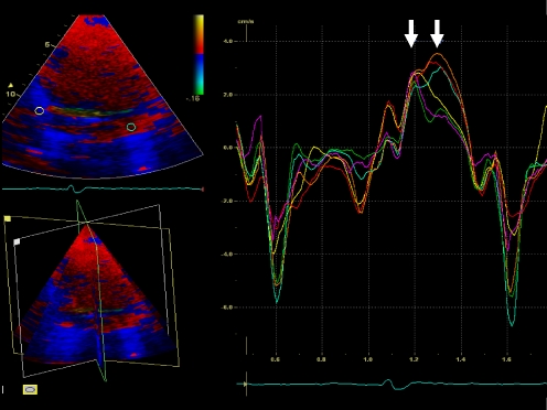
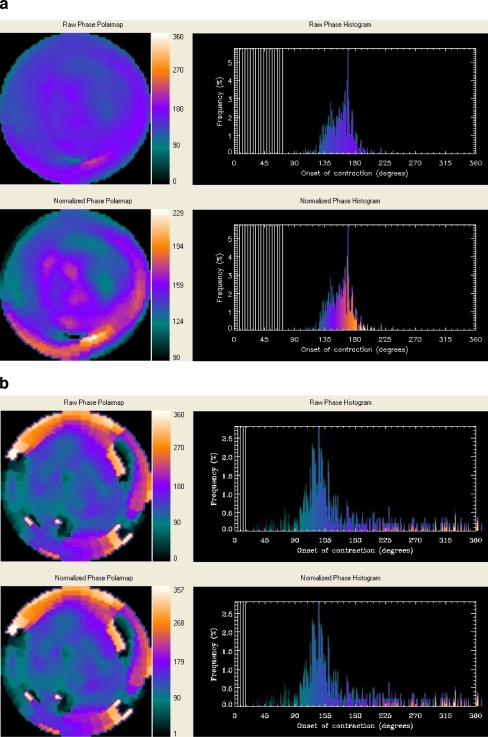
Purpose: To compare left ventricular (LV) dyssynchrony assessment by phase analysis from gated myocardial perfusion SPECT (GMPS) with LV dyssynchrony assessment by tri-plane tissue Doppler imaging (TDI). Baseline LV dyssynchrony assessed with standard deviation (SD) of time-to-peak systolic velocity of 12 LV segments (Ts-SD) with TDI has proven to be a powerful predictor of response to CRT. Information on LV dyssynchrony can also be provided by GMPS with phase analysis of regional LV maximal count changes throughout the cardiac cycle.

Methods: Forty heart failure patients, referred for evaluation of potential eligibility for CRT, underwent both 3D echocardiography, with tri-plane TDI, and resting GMPS. From tri-plane TDI, Ts-SD was used as a validated parameter of LV dyssynchrony and compared with different indices (histogram bandwidth, phase SD, histogram skewness and kurtosis) derived from phase analysis of GMPS.

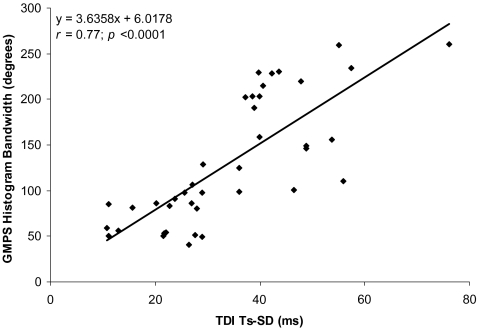
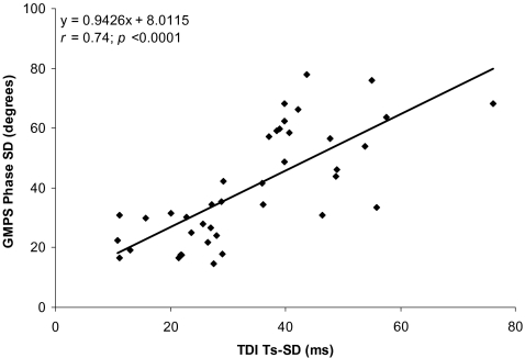
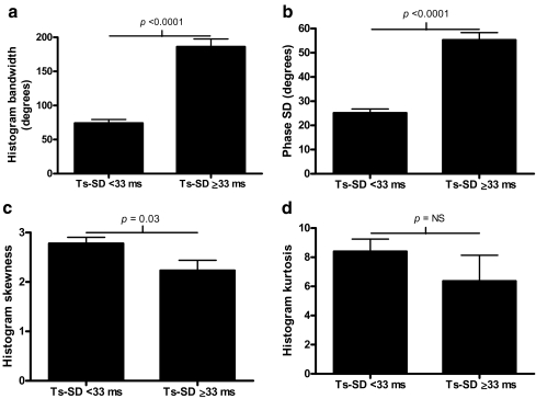
Results: Histogram bandwidth and phase SD showed good correlation with Ts-SD (r=0.77 and r=0.74, p<0.0001, respectively). Patients with substantial LV dyssynchrony assessed with tri-plane TDI (Ts-SD >or=33 ms) had also significantly higher values of histogram bandwidth and phase SD.

Conclusions: The results of this study support the use of phase analysis by GMPS to evaluate LV dyssynchrony. Histogram bandwidth and phase SD showed the best correlation with Ts-SD assessed with tri-plane TDI and appeared the most optimal variables for assessment of LV dyssynchrony with GMPS.


Figures

Fig. 1
Fig. 1
Example of the myocardial velocity curves that can be derived by positioning the sample volume in any LV segment of the tri-plane dataset. This patient has substantial LV dyssynchrony: the postero-lateral and anterior walls (orange, light blue and red curves) are activated later than the septal and inferior walls (yellow and green curves); standard deviation of 12 LV segments' Ts is 57.5 ms
Fig. 2
Fig. 2
a Example of a patient without LV dyssynchrony on GMPS. The non-normalized (upper panel) and normalized (lower panel) phase distributions are nearly uniform and the corresponding phase histograms are highly-peaked, narrow distributions. b Example of a patient (same as in Fig. 1) with LV dyssynchrony on GMPS. The non-normalized (upper panel) and normalized (lower panel) phase distributions show significant non-uniformity and the corresponding phase histograms have widespread distributions
Fig. 3
Fig. 3
Correlation between histogram bandwidth assessed with GMPS and LV dyssynchrony assessed with tri-plane TDI (Ts-SD)
Fig. 4
Fig. 4
Phase SD assessed with GMPS versus LV dyssynchrony assessed with tri-plane TDI (Ts-SD)
Fig. 5
Fig. 5
a Patients with substantial versus no substantial LV dyssynchrony (Ts-SD), using a cut-off value ≥33 ms. Histogram bandwidth is significantly higher in patients with substantial LV dyssynchrony. b Patients with substantial versus no substantial LV dyssynchrony (Ts-SD), using a cut-off value ≥33 ms. Phase SD is significantly higher in patients with substantial LV dyssynchrony. c Patients with substantial versus no substantial LV dyssynchrony (Ts-SD), using a cut-off value ≥33 ms. Histogram skewness is significantly lower in patients with substantial LV dyssynchrony. d Patients with substantial versus no substantial LV dyssynchrony (Ts-SD), using a cut-off value ≥33 ms. No significant difference in histogram kurtosis was demonstrated between patients without and patients with substantial LV dyssynchrony as assessed with tri-plane echocardiography

References

    1. Freemantle N, Tharmanathan P, Calvert MJ, Abraham WT, Ghosh J, Cleland JG. Cardiac resynchronisation for patients with heart failure due to left ventricular systolic dysfunction-a systematic review and meta-analysis. Eur J Heart Fail 2006;8:433–40. - DOI - PubMed
    1. Abraham WT, Fisher WG, Smith AL, Delurgio DB, Leon AR, Loh E, et al. Cardiac resynchronization in chronic heart failure. N Engl J Med 2002;346:1845–53. - DOI - PubMed
    1. Bax JJ, Bleeker GB, Marwick TH, Molhoek SG, Boersma E, Steendijk P, et al. Left ventricular dyssynchrony predicts response and prognosis after cardiac resynchronization therapy. J Am Coll Cardiol 2004;44:1834–40. - DOI - PubMed
    1. Sogaard P, Egeblad H, Kim WY, Jensen HK, Pedersen AK, Kristensen BO, et al. Tissue Doppler imaging predicts improved systolic performance and reversed left ventricular remodeling during long-term cardiac resynchronization therapy. J Am Coll Cardiol 2002;40:723–30. - DOI - PubMed
    1. Notabartolo D, Merlino JD, Smith AL, Delurgio DB, Vera FV, Easley KA, et al. Usefulness of the peak velocity difference by tissue Doppler imaging technique as an effective predictor of response to cardiac resynchronization therapy. Am J Cardiol 2004;94:817–20. - DOI - PubMed

Publication types

MeSH terms