. 2007 Sep 17:8:78.
doi: 10.1186/1471-2199-8-78.

GATA elements control repression of cardiac troponin I promoter activity in skeletal muscle cells

Affiliations

GATA elements control repression of cardiac troponin I promoter activity in skeletal muscle cells

Raffaella Di Lisi et al. BMC Mol Biol. .

Abstract

Background: We reported previously that the cardiac troponin I (cTnI) promoter drives cardiac-specific expression of reporter genes in cardiac muscle cells and in transgenic mice, and that disruption of GATA elements inactivates the cTnI promoter in cultured cardiomyocytes. We have now examined the role of cTnI promoter GATA elements in skeletal muscle cells.

Results: Mutation or deletion of GATA elements induces a strong transcriptional activation of the cTnI promoter in regenerating skeletal muscle and in cultured skeletal muscle cells. Electrophoretic mobility shift assays show that proteins present in nuclear extracts of C2C12 muscle cells bind the GATA motifs present in the cTnI promoter. However, GATA protein complex formation is neither reduced nor supershifted by antibodies specific for GATA-2, -3 and -4, the only GATA transcripts present in muscle cells.

Conclusion: These findings indicate that the cTnI gene promoter is repressed in skeletal muscle cells by GATA-like factors and open the way to further studies aimed at identifying these factors.


Figures

Figure 1
Figure 1
The cTnI promoter is activated by mutation or deletion of GATA sites in skeletal muscle in vivo. Transfection experiments of cTnI-CAT constructs in regenerating rat soleus muscles. The -230/+16 cTnI promoter and different mutants are schematically represented on the left, the three GATA site being indicated with squares and the A/T rich element with an oval. CAT activities in transfected muscles are shown on the right. The following mutants were examined (top to bottom): -145 and -127 deletion mutants, leading to the deletion of two of three or all three GA-rich sequences, respectively; mutation of the A/T-rich sequence (filled oval); GATA → CACA mutation of the proximal GATA site (crossed square); GATA → CACA mutation of the two distal GATA sites (two crossed squares); GATA → CACA mutation of all three GATA sites (three crossed squares); deletion of the two distal GATA sites; GATA → GCCA mutation of all three GATA sites (filled squares). Note that deletion/mutation of GATA sites leads to increased activity of the cTnI promoter, up to levels even higher than the SV40 promoter. Values (means ± S.E.) are expressed as fold increase in CAT activity over the promoterless construct. Each construct was tested in 4 to 8 independent experiments.
Figure 2
Figure 2
The cTnI promoter is activated by mutation or deletion of GATA sites in skeletal muscle cells in vitro. Transfection experiments of cTnI-CAT constructs in cultured primary cardiomyocytes (grey bars) and skeletal muscle cells (black bars). Schematic drawings of constructs are as in Fig. 1. Note that mutation of GATA sites leads to decreased promoter activity in cardiomyocytes but increased activity in skeletal muscle cells. Values (means ± S.E.) are expressed as fold increase in normalized CAT activity over the promoterless construct. Each construct was tested in 4 to 5 independent experiments.
Figure 3
Figure 3
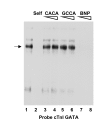
The cTnI promoter GATA motifs are target for a protein complex in nuclear extracts from C2C12 muscle cells. A 32P-labeled probe encompassing the two distal GATA elements in the cTnI promoter (see Table 1) shows a major retarded complex (arrow) when incubated with nuclear extracts from cultured C2C12 muscle cells (lane 1). For competition assays, the following non-radioactive competitors were added to the reaction mixture at 50- and 100- molar excess prior to the addition of the probe: self competitor (lane 2, 100-fold molar excess), CACA mutated cTnI GATA motifs (lanes 3 and 4), GCCA mutated cTnI GATA motifs (lanes 5 and 6), and a GATA sequence from the BNP promoter (lanes 7 and 8). The formation of the complex is prevented by the cTnI and BNP GATA sequences but is unaffected by the mutated cTnI GATA sequences.
Figure 4
Figure 4
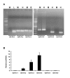
Muscle cells express GATA-2, -3 and -4 but not GATA-1, -5 and -6 transcripts. A. RT-PCR assays were performed with RNA preparations from differentiated C2C12 cells (M), using heart (H), liver (L) or brain (B) RNA preparations as positive controls. The primers used are indicated in the Method section. Note that muscle cells express GATA-2, -3 and -4 but not GATA-1, -5 and -6 transcripts. B. Relative mRNA expression levels of GATA transcripts were evaluated by quantitative real-time PCR, using cyclophilin A as an internal standard.
Figure 5
Figure 5
GATA-3 and -4 proteins are not detected and GATA-2 protein is barely detected by Western blotting in skeletal muscle cells. Western blotting was performed using antibodies specific for GATA-2, -3 or 4 and nuclear extracts of differentiated C2C12 cells (M) or control cells (He:Hela, J:Jurkat or H:heart cells). A control blot reacted with anti-MEF2 antibody demonstrates the presence of MEF2 in nuclear extracts of muscle cells. 20 μg of extract was loaded per lane.
Figure 6
Figure 6
The cTnI GATA elements do not bind GATA-2, -3 and -4 protein in muscle cell nuclear extracts. The 32P-labeled cTnI GATA probe was incubated with C2C12 nuclear extracts, Jurkat or cardiomyocyte nuclear extracts were used as controls. For supershift experiments, nuclear extracts were preincubated with anti-GATA-2, -3 or -4 antibody. In the presence of the anti-GATA-2 antibody, the major complex observed in the binding reaction of cTnI GATA probe with Jurkat nuclear extracts (arrow) is partially inhibited (compare lanes 1 and 2), while no inhibition is observed with the nuclear extract of C2C12 muscle cells (compare lanes 4 and 5). In the presence of the anti-GATA-3 antibody, a supershift is observed with Jurkat nuclear extract (compare lanes 1 and 3), but no supershift is seen with C2C12 nuclear extract (compare lanes 4 and 6). A supershift is also observed with cardiomyocyte nuclear extract in the presence of the anti-GATA-4 antibody (compare lanes 7 and 8), but no supershift is seen with C2C12 nuclear extracts (compare lanes 9 and 10).

Similar articles

Cited by

References

    1. Lee KJ, Hickey R, Zhu H, Chien KR. Positive regulatory elements (HF-1a and HF-1b) and a novel negative regulatory element (HF-3) mediate ventricular muscle-specific expression of myosin light-chain 2-luciferase fusion genes in transgenic mice. Mol Cell Biol. 1994;14:1220–1229. - PMC - PubMed
    1. Dhar M, Mascareno EM, Siddiqui MA. Two distinct factor-binding DNA elements in cardiac myosin light chain 2 gene are essential for repression of its expression in skeletal muscle. Isolation of a cDNA clone for repressor protein Nished. J Biol Chem. 1997;272:18490–18497. doi: 10.1074/jbc.272.29.18490. - DOI - PubMed
    1. Schiaffino S, Reggiani C. Molecular diversity of myofibrillar proteins: gene regulation and functional significance. Physiol Rev. 1996;76:371–423. - PubMed
    1. Di Lisi R, Millino C, Calabria E, Altruda F, Schiaffino S, Ausoni S. Combinatorial cis-acting elements control tissue-specific activation of the cardiac troponin I gene in vitro and in vivo. J Biol Chem. 1998;273:25371–25380. doi: 10.1074/jbc.273.39.25371. - DOI - PubMed
    1. Vitadello M, Schiaffino MV, Picard A, Scarpa M, Schiaffino S. Gene transfer in regenerating muscle. Hum Gene Ther. 1994;5:11–18. - PubMed

Publication types