. 2007 Sep 15;21(18):2300-11.
doi: 10.1101/gad.1564307.

Drosophila PIWI associates with chromatin and interacts directly with HP1a

Affiliations

Drosophila PIWI associates with chromatin and interacts directly with HP1a

Brent Brower-Toland et al. Genes Dev. .

Abstract

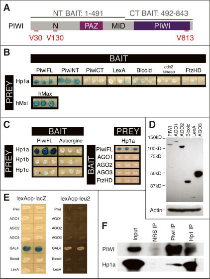
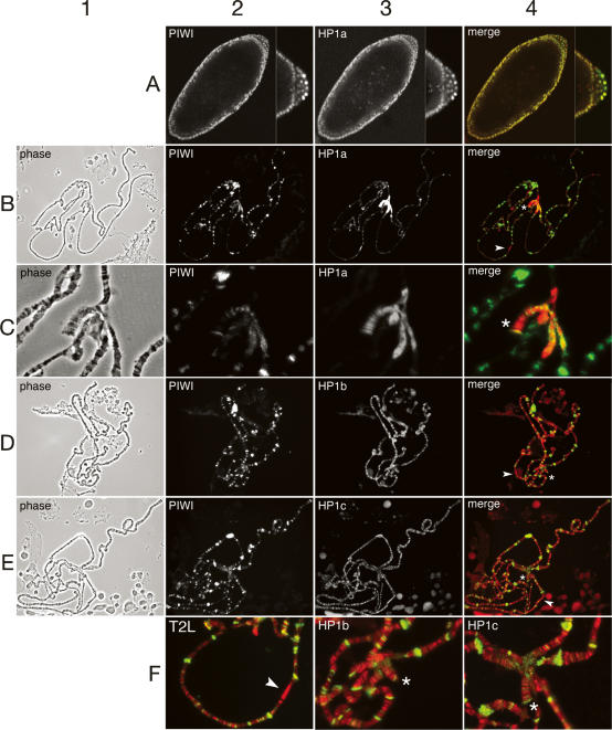
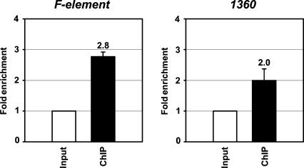
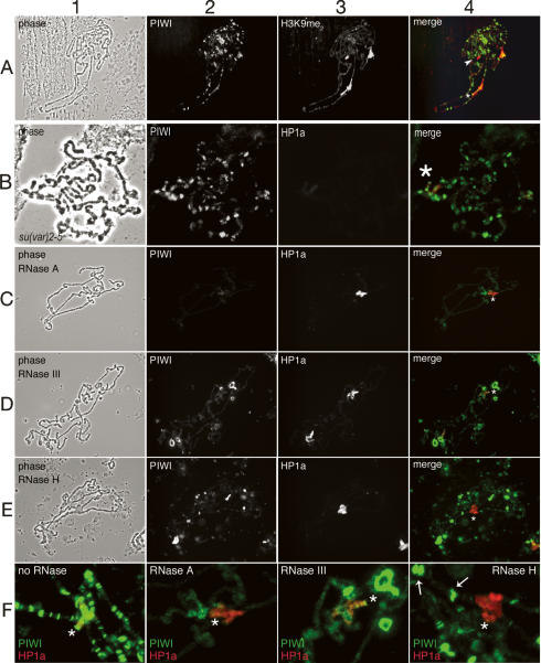
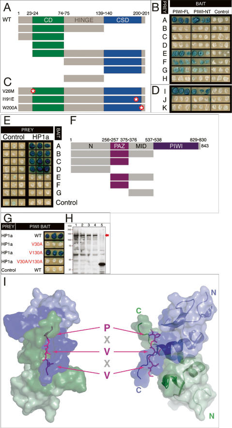
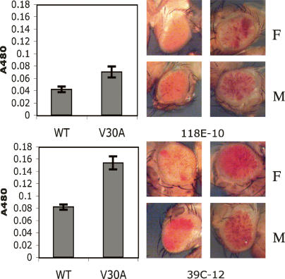
The interface between cellular systems involving small noncoding RNAs and epigenetic change remains largely unexplored in metazoans. RNA-induced silencing systems have the potential to target particular regions of the genome for epigenetic change by locating specific sequences and recruiting chromatin modifiers. Noting that several genes encoding RNA silencing components have been implicated in epigenetic regulation in Drosophila, we sought a direct link between the RNA silencing system and heterochromatin components. Here we show that PIWI, an ARGONAUTE/PIWI protein family member that binds to Piwi-interacting RNAs (piRNAs), strongly and specifically interacts with heterochromatin protein 1a (HP1a), a central player in heterochromatic gene silencing. The HP1a dimer binds a PxVxL-type motif in the N-terminal domain of PIWI. This motif is required in fruit flies for normal silencing of transgenes embedded in heterochromatin. We also demonstrate that PIWI, like HP1a, is itself a chromatin-associated protein whose distribution in polytene chromosomes overlaps with HP1a and appears to be RNA dependent. These findings implicate a direct interaction between the PIWI-mediated small RNA mechanism and heterochromatin-forming pathways in determining the epigenetic state of the fly genome.


Figures

Figure 1.
Figure 1.
HP1a interacts specifically with PIWI. (A) Baits used in PIWI Y2H screens. PIWI contains N, PAZ, MID, and PIWI domains. Y2H baits are PIWI-FL, residues 1–843; PIWI-NT, residues 1–491; and and PIWI-CT, residues 492–843. The positions of the three “PxV” sequences—at V30, V130 and V813—are indicated. (B) HP1a is a strong PIWI interactor. Three independent transformants for each bait/prey combination were plated to assess LacZ activity of reporter plasmid pSH18-34. HP1a interacts strongly with PIWI-FL and PIWI-NT baits, but not with PIWI-CT or unrelated baits, including the LexA moiety of the bait plasmid pEG202, Drosophila BICOID, Drosophila Cdc2 kinase or Drosophila FUSHI-TARAZU homeodomain. The strongly interacting human Mxi and Max proteins serve as a positive control. (C) The PIWI–HP1a interaction is specific. Full-length PIWI interacts with HP1a, but not with closely related HP1b or HP1c. Similarly, HP1a shows no interaction with PIWI paralogs AUB, AGO1, AGO2, or AGO3. (D) AGO1 is expressed at a similar level to PIWI, whereas AGO2 and AGO3 are expressed much more abundantly than PIWI, as shown by the Western blot analysis. (E) AGO1, AGO2, and AGO3 proteins enter the nucleus, as shown by lexA–lacZ reporter and lexA–leu2 expression-blocking assays (see Materials and Methods). (F) PIWI and HP1a interact in vivo. Nuclear proteins from Drosophila embryos were incubated with antibodies specific for HP1a or PIWI and the resulting immunoprecipitates were analyzed for coprecipitation by Western blot analysis. For reference, HP1a and PIWI immunoprecipitates are compared with material generated using naive rabbit serum (NRS).
Figure 2.
Figure 2.
PIWI is associated with chromatin, where it colocalizes with HP1a. All images are from wild-type (Oregon R) embryos or larval salivary glands. In merged images showing PIWI (green channel) and HP1 isoforms (red channel), the overlap is yellow. On merged images, arrowheads indicate cytological region 31; the fourth chromosome is denoted by an asterisk. (A) PIWI colocalizes with HP1 in all nuclei of stage 5 embryos; PIWI is more abundant in pole cell nuclei (posterior pole is shown at higher magnification at right of each embryo image). (B) PIWI shows extensive colocalization with HP1a to euchromatic bands along polytene chromosome arms, to telomeres, and to distinct regions of the chromocenter. HP1a is most concentrated in the chromocenter and along chromosome 4. (C) PIWI partially colocalizes with HP1a in the chromocenter and at distinct bands on the largely heterochromatic chromosome 4. (D–E) PIWI localization is distinct from and largely nonoverlapping with HP1b and HP1c. PIWI does not overlap with HP1b or HP1c in region 31. (F) Enlargements from merged images in C–E, emphasizing that PIWI colocalizes with HP1a at telomere 2L, is not concentrated in region 31A, and is distinct from both HP1b and HP1c in the chromocenter.
Figure 3.
Figure 3.
PIWI is enriched in the HP1a-rich F and 1360 elements. Shown are the ratios of the abundance of PIWI-associated DNA of F and 1360 elements versus rpL32 DNA obtained by MYC-PIWI immunoprecipitation of cross-linked chromatin. The results represent the average values of six independent samples.
Figure 4.
Figure 4.
PIWI chromatin binding overlaps H3K9 methylation and depends on RNA hybrids but not HP1a. Images from indirect immunofluorescent detection of PIWI and HP1a. Labels and color channels are as in Figure 3. All images are of wild-type (Oregon R) chromosomes, except B, which shows Su(var)2-5 mutant chromosomes. (A) PIWI shows significant overlap with dimethyl-H3K9 in the chromocenter and fourth chromosome but not region 31 (arrowhead). (B) PIWI localization to polytene chromosomes is not globally perturbed in the absence of HP1a. Su(var)2-5 chromosomes are shown at higher magnification because of their small size by comparison with wild-type chromosomes. (C–F) Patterns of PIWI and HP1a (determined by indirect immunofluorescence) on chromosomes treated with different RNase activities. In each case, the HP1a-binding pattern remains essentially the same as on untreated chromosomes. On all merged images the fourth chromosome is indicated by an asterisk. (C) Mild treatment with RNase A eliminates the PIWI-binding pattern seen on untreated chromosomes without perturbing HP1a. (D) RNase III treatment results in loss of PIWI binding to bands in the euchromatic arms without abolishing PIWI bound in pericentric chromatin and in bands on chromosome 4. Delocalized PIWI is concentrated in ring-like bodies that may represent nucleolar fragments in the polytene preparation. (E) PIWI binding in pericentric chromatin and chromosome 4 is abolished by RNase H digestion; binding along the euchromatic arms is diminished at some sites more than others, but not abolished. (F) Enlargement of chromocenters from merged images in C–E with a representative image from untreated chromosomes for comparison. Arrows point to the brightly staining PIWI bands in euchromatic arms, not affected by RNase H treatment.
Figure 5.
Figure 5.
HP1a requires an intact CSD dimer interface to bind PIWI. (A) PIWI requires an intact CSD dimer interface to bind HP1a. Cartoon of the HP1a deletion series used to map PIWI-binding requirements. The 206-residue HP1a contains a CD, a hinge domain, and a CSD. Domain junction residue numbers are indicated (WT, full-length, wild type). (B) Only prey possessing the CSD (prey A, E, F, and G) interact with PIWI-FL and PIWI-NT baits. (C,D) PIWI interaction is lost with HP1a mutations predicted to disrupt either its target binding interface within the intact CSD dimer (W200A mutant) or dimerization (191E mutant), but not with a mutation in the CD (V26M). Positions of the respective point mutations are indicated by stars. (E) Only baits containing the N domain of PIWI (baits A–D) interact with HP1a-FL. (F) Cartoon of the PIWI deletion series used to map HP1a binding requirements. Domain junction residue numbers are indicated. (G) V30 of PIWI but not V130 is required for HP1a interaction. Wild type and V130A PIWI mutant produce comparably strong LacZ signals, whereas the signal in V30A or the V30A/V130A double mutant is undetectable. (H) Western blot of protein extracts of yeast strains in G. Each bait is expressed at comparable levels, as detected by polyclonal antibody to LexA, the DNA-binding moiety common to each bait. (Lane 1) Wild-type PIWI. (Lane 2) V30A. (Lane 3) V130A. (Lane 4) V30A/V130A. (Lane 5) BICOID homeodomain. The LexA-PIWI baits (red arrow) are predicted to be 120.2 kDa. (I) Model of the HP1a CSD dimer in complex with the PIWI peptide (TSRGSGDGPRVKVFRGSSSGD). Top and side views are shown in left and right panels, respectively. Each monomer of the HP1a CSD dimer is color-coded (green or blue). The CSD-binding motif (PxVxV) in the PIWI peptide is shown in stick models (magenta). Side chains of the conserved residues (P,V,V) are displayed. Chemical shift perturbations are calculated as δCS = [δH2 + (0.2δN)2]1/2. Residues of HP1a that experience significant resonance perturbation (δCS > 0.06 ppm) during PIWI peptide titration are colored in dark green and dark blue and are mapped on the CSD surface and the ribbon diagram. The homology model of the complex was built using the XLOOK program (Lee 1993), and the figure was generated by PyMOL (Delano Scientific).
Figure 6.
Figure 6.
A PIWI transgene bearing the V30A mutation is deficient in rescuing dominant defects in white reporter silencing produced by the piwi2 mutation. Eye pigmentation of PIWIWT; piwi2/CyO;P[wvar] flies are compared with pigmentation in PIWIV30A;piwi2/CyO;P[wvar] flies. Variegating white reporters (P[wvar]) analyzed are embedded in pericentric (118E-10) or fourth chromosome (39C-12) heterochromatin. (F) Female; (M) male.

References

    1. Alfageme C.R., Rudkin G.T., Cohen L.H., Rudkin G.T., Cohen L.H., Cohen L.H. Locations of chromosomal proteins in polytene chromosomes. Proc. Natl. Acad. Sci. 1976;73:2038–2042. - PMC - PubMed
    1. Bartels C., Xia T., Billeter M., Güntert P., Wüthrich K., Xia T., Billeter M., Güntert P., Wüthrich K., Billeter M., Güntert P., Wüthrich K., Güntert P., Wüthrich K., Wüthrich K. The program XEASY for computer-supported NMR spectral analysis of biological macromolecules. J. Biol. NMR. 1995;6:1–10. - PubMed
    1. Brasher S.V., Smith B.O., Fogh R.H., Nietlispach D., Thiru A., Nielsen P.R., Broadhurst R.W., Ball L.J., Murzina N.V., Laue E.D., Smith B.O., Fogh R.H., Nietlispach D., Thiru A., Nielsen P.R., Broadhurst R.W., Ball L.J., Murzina N.V., Laue E.D., Fogh R.H., Nietlispach D., Thiru A., Nielsen P.R., Broadhurst R.W., Ball L.J., Murzina N.V., Laue E.D., Nietlispach D., Thiru A., Nielsen P.R., Broadhurst R.W., Ball L.J., Murzina N.V., Laue E.D., Thiru A., Nielsen P.R., Broadhurst R.W., Ball L.J., Murzina N.V., Laue E.D., Nielsen P.R., Broadhurst R.W., Ball L.J., Murzina N.V., Laue E.D., Broadhurst R.W., Ball L.J., Murzina N.V., Laue E.D., Ball L.J., Murzina N.V., Laue E.D., Murzina N.V., Laue E.D., Laue E.D. The structure of mouse HP1 suggests a unique mode of single peptide recognition by the shadow chromo domain dimer. EMBO J. 2000;19:1587–1597. - PMC - PubMed
    1. Brennecke J., Aravin A.A., Stark A., Dus M., Kellis M., Sachidanandam R., Hannon G.J., Aravin A.A., Stark A., Dus M., Kellis M., Sachidanandam R., Hannon G.J., Stark A., Dus M., Kellis M., Sachidanandam R., Hannon G.J., Dus M., Kellis M., Sachidanandam R., Hannon G.J., Kellis M., Sachidanandam R., Hannon G.J., Sachidanandam R., Hannon G.J., Hannon G.J. Discrete small RNA-generating loci as master regulators of transposon activity in Drosophila. Cell. 2007;128:1089–1103. - PubMed
    1. Caudy A.A., Myers M., Hannon G.J., Hammond S.M., Myers M., Hannon G.J., Hammond S.M., Hannon G.J., Hammond S.M., Hammond S.M. Fragile X-related protein and VIG associate with the RNA interference machinery. Genes & Dev. 2002;16:2491–2496. - PMC - PubMed

Publication types

MeSH terms